Cell adhesion molecules, Cell junctions types, definition and function
Intercellular junctions are structures that provide adhesion & communication between cells. They are mostly present in epithelial cells that are especially characterized by their strong attachment one to another and to the extracellular matrix but can also exist between other types of cells e.g. cardiac muscle fibers.
Cell adhesion molecules
Cells adhere either directly to each other in cell-to-cell adhesion or to extracellular matrix in cell-to-matrix adhesion, certain glycoprotein molecules mediate cell adhesion and they are called cell adhesion molecules (CAMs). Cell adhesion molecules are transmembrane glycoproteins, each consists of three domains:
- Extracellular (EC) domain binds with the other CAMs of adjacent cells or extracellular matrix proteins.
- Intramembranous (transmembrane) domain.
- The cytoplasmic domain is attached to the cytoskeleton of the cell through linker Proteins.
The main CAMs include integrins, cadherins, selectins, and immunoglobulin superfamily, CAMs may be Ca+2-dependant i.e. affected by the extracellular calcium ions concentration, whereas others are Ca+2 independent.
- Integrins are Ca+2-dependent CAMs and they are involved mainly in cell-to-extracellular matrix adhesion.
- Cadherins are Ca+2-dependent CAMs and they are involved in cell-to-cell adhesion. Selectins are Ca+2-dependent CAMs and they are involved in cell-to-cell adhesion.
- Immunoglobulin superfamily: the molecules of this family are Ca+2– independent CAMs and they are involved in cell-to-cell adhesion, they are a large family that includes; vascular cell adhesion Molecule (V-CAM) and neural cell adhesion molecule (N-CAM) which plays an important role in nervous system development.
Cell adhesion is a dynamic process that is particularly evident where non-adhesive cells rapidly become adhesive e.g. leukocytes in inflammation and platelets in blood clotting, the reverse takes place where the temporal loss of adhesion occurs during the migration of skin cells in wound healing to replace lost cells, on the other hand, defective cellular adhesion in malignancy promotes spread of cancer cells and occurrence of metastasis.
Types of connecting cell junctions
There are 3 types of connecting cell junctions; occluding, anchoring, and communicating junctions:
Occluding junction (tight junction, zonula occludens)
This junction is present at the apical parts of the cells, Zonulae occludentes are found mainly where no substance should pass in the intercellular space e.g. simple columnar epithelial cells lining the intestine to prevent the passage of the digestive juices and the bacteria to the intercellular spaces. Occluding junction restricts the passage of water, electrolytes, and other small molecules between the epithelium, thereby contributing to the barrier function of the cells.
Histological structure
- The outer leaflets of adjacent cell membranes are fused together, around the whole perimeter of each cell, forming a continuous belt-like junction (zonula = Latin for belt)
- 2 transmembrane proteins, occludin and claudin are embedded in each of the two adjacent cell membranes; join directly to one another to produce a tight seal and to occlude the intercellular space i.e. no intercellular space between the opposing cell membranes.
Anchoring junctions
They are most widely distributed in cells that are subjected to severe mechanical stress such as cardiac muscles and the epidermis of the skin. They provide strong attachments and act as a link between the cytoskeleton of adjacent cells.
Histological structure
Two types of anchoring junctions can be identified:
1- Zonula adherens:
- It is a belt-like specialization of the cell membrane and the subjacent cytoplasm that encircles the apical part of the 2 adjoining cells.
- The intercellular space between the opposing cell membranes is 20 nm (this is the usual intercellular space between adjacent epithelial cells).
- Cadherins interact to adhere the two cells together.
- The cytoplasmic components of cadherins are attached to actin filaments inside the cells while their extracellular domains are linked in the presence of Ca+2, Removal of Ca+2 leads to disruption of the zonula.
2- Macula adherens (the desmosome):
- It is a spot-like specialization of the cell membrane and the subjacent cytoplasm, in which the intercellular space between the opposing cell membranes is 30 nm.
- Cadherins interact to connect the two cells together.
- Desmosome is composed of 2 disk-shaped attachment plaques located opposite each other on the cytoplasmic aspects of the cell membranes of adjacent cells, to which cytokeratin intermediate filaments are inserted. Dense vertical filamentous material is present in the intercellular space between the opposing cell membranes that represents the extracellular domains of cadherins, which link the cells firmly with the help of calcium ions.
- In the presence of a calcium-chelating agent, the desmosome breaks into 2 halves, and the cells separate.
In several epithelia, the zonula occludens, the zonula adherens and macula adherens are present in a definite order from the apex toward the base of the cell and is called intercellular junctional complexes.
Hemidesmosomes (basal cell-to-matrix adhesions):
- They look like half of the desmosome but are different functionally and in their content.
- Hemidesmosomes are found at the base of the epithelial cells to connect them with the underlying basement membrane.
- The CAMs are not cadherins but integrins; their extracellular domains bind to proteins of the basal lamina while the intracellular domains bind to cytokeratin intermediate filaments.
Communicating junction (gap junction, nexus)
They mediate communication rather than adhesion and beside epithelial cells are also found in cardiac and smooth muscle cells. They permit the exchange of molecules e.g. ions, amino acids allowing passage of signals involved in contraction, integration, and communication from one cell to another.
Histological structure:
- It is a spot-like junction, formed of protein channels that communicate the interior of two adjacent cells; the intercellular space in this junction is narrowed to about 3nm.
- It is formed of the connexons which are formed of 6 closely-packed transmembrane proteins, the connexins; not attached to specific cytosolic filaments.
- When two connexons of opposing cell membranes are in register, they form a channel connecting the cytoplasm of adjacent cells.
Epithelial polarity, Apical, basal & lateral surfaces of epithelial cells
Tissues types, Epithelial tissue features, Covering & Glandular Epithelium
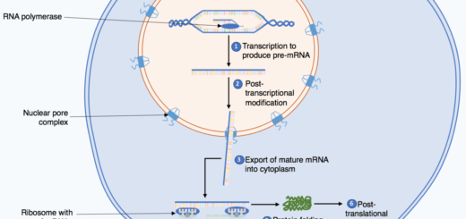
DNA Repair types, definition & importance, Protein Biosynthesis & Genetic Code
Protein biosynthesis steps, site, importance, inhibitors & Protein maturation