Histological organization of spinal cord, Relation between spinal and vertebral segments
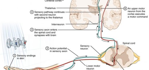
The spinal cord carries signals from the brain: It receives signals from the brain that control movement and autonomic functions, It carries information to the brain: The spinal cord nerves also transmit messages to the brain from the body, such as sensations of touch, pressure, and pain, The spinal cord may act independently of the brain in conducting motor reflexes.
Spinal cord
Histologically, the CNS is formed of grey and white matter. The grey matter, its color in the fresh state is grey. It is composed of:
- Nerve cell bodies,
- Nerve fibers (mostly unmyelinated),
- A network of neuroglia,
- A rich capillary bed.
Nerve cells in the grey matter are aggregated into large and small nuclei. A nucleus in CNS: it is a group of nerve cells lying close to each other and having the same function. The white matter, its color in the fresh state is white. It is composed of:
- Bundles of nerve fibers (mostly myelinated),
- A network of neuroglia.
- Fewer blood capillaries.
Myelinated nerve fibers in the white matter are longitudinally-arranged in ascending and descending tracts. A tract in CNS: it is a group of nerve fibers having the same origin and termination and carrying the same function.
Histological organization of spinal cord
The spinal cord is an elongated cylindrical cord, about 45 cm long, and occupies the upper 2/3 of the vertebral canal. It starts at the upper border of C1 vertebra and ends at the lower border of L1 vertebra. The spinal cord is differentiated into 31 segments: 8 cervical, 12 thoracic, 5 lumbar, 5 sacral, and 1 coccygeal.
The spinal cord presents two enlargements:
- Cervical enlargement extends from C3 to T2 and corresponds to the origin of the brachial plexus.
- Lumbosacral enlargement extends from L1 to S3 and it corresponds to the origin of the lumbar and sacral plexuses.
Relation between spinal and vertebral segments
- The spinal cord occupies the whole length of the vertebral canal up to the 3rd month of intra-uterine life.
- As age advances, the vertebral column grows faster than the spinal cord (differential growth), the spinal cord ends at the level of L3 vertebra at birth.
- In adults, the lower end of the cord lies opposite the lower border of the L1 vertebra, The dura and arachnoid maters end at the level of the S2 vertebra, Therefore, the spinal segments do not lie opposite the corresponding vertebrae.
A transverse section shows that the spinal cord is a bilateral structure, consisting of central grey matter and peripheral white matter. The two halves are separated posteriorly by the posterior median septum and anteriorly by the anterior median fissure. They are continuous in the intermediate zone through the grey and white commissures. The central canal is always present in the middle of the grey commissure.
Grey matter of the spinal cord
Unlike the cerebrum and cerebellum, the grey matter in the spinal cord is central in position. On each side, the grey matter is divided into three horns, also called grey columns. These are; the ventral horn, the dorsal horn, and the lateral horn, whose shapes and sizes differ in different regions of the spinal cord.
Laminar architecture of spinal cord: the grey matter of the spinal cord is divided into 10 laminae (from I to X) based upon the cytological features of the neurons in different regions of grey matter.
Nuclei in spinal cord grey matter
1. The ventral horn (anterior horn) (motor nuclei)
This horn is broad in the cervical and lumbosacral segments (a greater amount of grey matter for the innervation of muscles of upper and lower limbs). It contains large stellate lower motor neurons (the anterior horn cells). Their axons form the motor fibers emerging in the ventral root of spinal nerves.
Neurons of the ventral horn are divided into 3 groups of nuclei:
- The medial group: it is further subdivided into the ventro-medial, and the dorso-medial nuclei which are represented in all segments of the spinal cord.
- The central group: it is only present in the cervical and lumbosacral enlargements. In the cervical region, it is present from C3 to C5 and is termed the phrenic nucleus.
- The lateral group: it is further subdivided into the ventro-lateral, and the dorso-lateral nuclei. They are like the central group i.e. present in the cervical, lumbar, and sacral segments, but absent in the thoracic region.
2. The dorsal horn (posterior horn) (sensory nuclei)
The cells of the dorsal horn are small stellate neurons (afferent neurons) that belong to the sensory system (they receive and process sensory input). They are grouped into 3 main nuclei:
- The substantia gelatinosa of Rolando: it is associated with the sensory pathway of slow pain and temperature and shares in the formation of the anterolateral system. It is present at the tip of the dorsal horn and is represented in all segments of the spinal cord.
- The nucleus proprius (main sensory nucleus): it is associated with the sensory pathway of fast pain and shares in the formation of the anterolateral system. lt lies in the central part of the horn and is represented in all segments of the spinal cord.
- The nucleus dorsalis of Clarke (nucleus thoracicus) (Clarke’s column): It is associated with the unconscious proprioceptive sensation and gives rise to the “dorsal” spinocerebellar tract. It lies at the base of the dorsal horn only from T1 to L3 spinal segments).
3. The lateral horn (intermediate grey matter) (sympathetic nuclei
This horn appears only from T1 to L2 segments). It is found between the dorsal and ventral horns. The cells are small stellate neurons, belonging to the autonomic nervous system (thoraco-lumbar outflow); their axons form the preganglionic sympathetic nerve fibers. In S2, S3 & S4, the autonomic neurons in the intermediate grey matter give the preganglionic parasympathetic outflow without forming a lateral horn.
Associative nuclei (interneurons are found in all the grey matter (anterior and posterior horns); they are formed of small nerve cells with short axons and are essential in local spinal cord function. They are of two types:
- Commissural neurons, their axons cross to the opposite side.
- Intersegmental neurons, their axons ascend and descend for a few segments on the same side.
White matter of the spinal cord
The white matter occupies the peripheral region of the spinal cord. It is subdivided by the two horns of the grey matter in each half into three columns. These are; the dorsal white column, the ventral white column, and the lateral white column.
- The ventral (anterior) white column: between the anterior median fissure and the anterior horn at the exit of the ventral root of the spinal nerve.
- The dorsal (posterior) white column: between the posterior median septum and the posterior horn. In the cervical and upper thoracic regions, this column is further divided by the posterior intermediate septum into the gracile tract medially and cuneate tract laterally.
- The lateral white column: lateral to both posterior and anterior horns.
Tracts of the spinal cord white matter are classified into; short and long tracts:
I. Short tracts
They begin and end within the spinal cord originating from the associative nuclei, thus they are associative in function.
II. Long tracts
1. Ascending (sensory) tracts arise from the nerve cells in the spinal ganglia or from the nerve cells of the posterior horns, and end in higher sensory centers. They are of two types:
a. Tracts carrying sensations that will finally reach the Cerebral cortex. They include:
- Gracile & cuneate tracts (Dorsal column pathway).
- The ventrolateral pathways (spinothalamic, spinoreticular, and spinomesencephalic tracts).
b. Tracts carrying impulses that will not reach the cerebral cortex but end in the subconscious level, the cerebellum or midbrain. They include:
- Dorsal, ventral spinocerebellar & cuneocerebellar tracts.
- Spino-olivary tract.
- Spino-tectal tract.
2. Descending (motor) tracts begin in higher motor centers in the cerebral cortex and brainstem and end in the anterior and lateral horns of the spinal cord. They include:
a. Pyramidal tracts: The ventral corticospinal tract (uncrossed pyramidal tract) and the lateral corticospinal tract crossed pyramidal tract).
b. Extra-pyramidal tracts:
- Rubrospinal tract.
- Lateral and medial (ventral) reticulospinal tracts.
- Tectospinal tract.
- Lateral and medial (ventral) vestibulospinal tracts.
The transverse section of the spinal cord at its different levels is correlated to the shape and size of grey and white matter in these levels.
You can follow science online on Youtube from this link: Science online
You can download Science online application on google Play from this link: Science online Apps on Google Play
Physiology of central human reflexes, Types & properties of Spinal cord reflexes
Physiology & function of the spinal cord, Lateral & medial brainstem pathway
Features of synaptic transmission, Mechanism of sensitization & long term potentiation
Synaptic transmission steps, Synapses types & Nature of the postsynaptic change
Blood supply of CNS (central nervous system), Carotid system & Circle of Willis
Skull function, anatomy, structure, views & Criteria of neonatal skull