Nervous tissues function, structure, types of Neurons and Nervous fibers
Nervous tissues consist of different types of neurons, all have an axon, they are called neural tissues, Nervous tissue is the main component of the nervous system. The nervous system regulates and controls body functions and activity. The nervous system consists of two parts: the central nervous system (CNS), and the peripheral nervous system (PNS).
Nervous tissue
It is one of the four primary basic tissues from which the body is made. It consists of two types of cells; neurons (nerve cells) and neuroglia (supporting cells).
Neurons
The neuron or nerve cell is the structural and functional unit of the nervous tissue.
General characteristics of neurons:
- Excitability as they respond to environmental changes by the generation of action potential or nerve impulse.
- Conductivity as they are capable of propagation of nerve impulses to other neurons, muscles, and glands.
Histological structure of the neuron:
It is composed of a cell body called the perikaryon or the soma and cull processes that include axon and dendrites.
1- The cell body: it is the part of the neuron containing the nucleus. It is composed of:
A. Cytoplasm: It contains organelles and inclusion, particularly:
- Nissl bodies (Nissl substance): by LM, they appear as basophilic granules. By EM, they are aggregates of ribosomes and rER, for protein synthesis. They are distributed in the cell body except in the region of the axon hillock.
- Larg perinuclear Golgi apparatus for packaging of neurotransmitters into synaptic vesicles.
- The cytoskeleton formed of neurofibrils that include neurofilaments and microtubules playing a role in the transmission of nerve impulses.
- Inclusions as lipofuscin pigments lipids.
B. Nucleus: neuron is metabolically-active and hence the nucleus is euchromatic.
2- Nerve cell processes
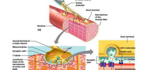
A. The axon:
- Origin: arises from the axon hillock.
- Number and shape: always single. Axon is long. with a constant diameter having a regular cylindrical shape.
- Branching: no branches except at axon termination forming terminal arborizations. It may give off collaterals arising at right angles.
- Histological structure: the axoplasm contains few organelles (neurofibrils, synaptic vesicles, and mitochondria). Nissl bodies are absent.
- Surrounding sheath: axolemma may be surrounded by sheaths according to the type of nerve fiber.
- The direction of impulses: conducts nerve impulses away from the cell body.
B. The dendrite:
- Origin: arises from any part of the cell body.
- Number and shapes: usually multiple (in multipolar neurons). It may be single (in bipolar neurons). Dendrite is short, thick near its origin, and tapers as it goes towards its end.
- Branching: many branches arising at acute angles, having short spines for synapses.
- Histological structure: contains most of the organelles as in the perikaryon except the Golgi apparatus. Nissl bodies are present.
- Surrounding sheath: not surrounded by sheaths.
- The direction of impulses: conducts nerve impulses towards the cell body.
Classification of the neurons
Functionally, neurons could be:
- Sensory neurons carry impulses from receptors to the central nervous system (CNS).
- Motor neurons carry impulses from CNS to the effector organs.
- Interneurons (association neurons) act as a link between sensory and motor neurons in CNS only.
Morphologically, neurons could be classified according to the number of their processes into:
- Unipolar neurons: have only one cell process, present in the embryonic stage.
- Pseudounipolar neurons have a single process that divides like the letter T into two branches (both are axons). They are the sensory neurons present in the cranio-spinal ganglia.
- Bipolar neurons: have two processes, one is an axon and the other is a dendrite. They are like the olfactory neurons present in the olfactory mucosa of the nose.
- Multipolar neurons: have more than two processes, These are further classified according to the shape of their perikaryon into:
- Stellate neurons are the anterior horn cells of the spinal cord and the autonomic ganglion cells.
- Pyramidal neurons present in the cerebral cortex.
- Pyriform neurons present in the cerebellar cortex (Purkinje cells) and in the retina (ganglion cells).
- Granule cells present mainly in the cerebellar cortex.
General organization of the nervous system
In the nervous system the cell bodies and their processes have the following distribution:
In the central nervous system (CNS); which comprises the brain and spinal cord, the cell bodies are present mainly in the grey matter while their axons (nerve fibers) are present mainly in the white matter.
In the peripheral nervous system (PNS); which includes nerve endings, peripheral nerves (somatic & autonomic), and the ganglia (cranio-spinal & autonomic). The neuronal cell bodies are present mainly in the ganglia while the axons (nerve fibers) are forming the peripheral nerves.
Nervous fibers
A nerve fiber consists of an axon enveloped by a special sheath. Nerve fibers exhibit differences in their enveloping sheaths, related to whether the fibers are part of the central or peripheral nervous system.
Types of nerve fibers
I. In peripheral nervous system:
- Myelinated with myelin sheath and neurilemma (formed by the neuroglia cells of the PNS, the Schwann cells). They are present in the peripheral nervous (nerve trunks).
- Unmyelinated with neurilemma only. They are present mainly in the autonomic nervous system.
- Naked uncovered by the sheath, present in the nerve endings (terminal part of the peripheral nervous, including the receptors and the effectors).
A. Myelinated nerve fibers in PNS: They are axons of large diameters and are enveloped by a myelin sheath and neurilemmal sheath.
I. The myelin sheath: It is a lipid-rich coat that covers the axon. It is formed by wrapping of Schwann cell around the axon. It is interrupted at equal intervals by constrictions (nodes of Ranvier) where the axon can emit its collaterals. The segment between two nodes is the internodal segment and is occupied by a single Schwann cell. At each node, the outer Schwann cell sheath meets the axon providing it with nutrients, as it is not covered by myelin.
Function: Insulation and protection of the nerve fiber as well as fast conduction of nerve impulse
II. Schwann cell sheath (neurilemmal sheath or neurilemma): It consists of the Schwann cells (cytoplasm and nucleus) that form an outer thin sleeve around the myelin of a nerve fiber.
Function: Formation of myelin, nutrition of the axon (at nodes of Ranvier) as well as regeneration of the nerve fiber after injury.
B- Unmyelinated nerve fibers in PNS: Axons are usually small in diameters; they are enveloped by Schwann cell sheath (neurilemma) only. A single Schwann cell envelops multiple segments of different axons.
II. In central nervous system:
Myelinated with myelin sheath (formed by neuroglia cells of the CNS, oligodendrocytes). They are present in white matter and the optic nerve. Naked nerve fibers are present in grey matter.
Myelinated nerve fibers in the CNS: Nerve fibers are enveloped by myelin sheath without neurilemma. Here, oligodendrocytes differ from Schwann cells, which are absent in CNS in that multiple processes of single oligodendrocyte can envelop segments of several nearby axons. The cell body of the oligodendrocyte will be at some distance from the axons it myelinates.
Flexors of forearm, Forearm muscles, structure, function & anatomy
Forearm bones, anatomy, function & Skeleton of the hand
Arm structure, compartments, muscles, anatomy & Cubital Fossa contents
Bones of upper limb structure, function, types & anatomy