Hands structure, function, bones, nerves, muscles and anatomy
The hand is a prehensile, multi-fingered appendage located at the end of the forearm or forelimb of primates such as humans, chimpanzees, monkeys, and lemurs. The human hand has five digits: four fingers plus one thumb; these are referred to collectively as five fingers, however, whereby the thumb is included as one of the fingers.
Hand
The hand has 27 bones, not including the sesamoid bone, the number of which varies among people, 14 of which are the phalanges (proximal, intermediate and distal) of the fingers and thumb. Fingers contain some of the densest areas of nerve endings in the body and are the richest source of tactile feedback. the hand is responsible for the creative manifestations that characterize the human species and that distinguish it from all other known forms of life.
Cutaneous nerve supply
Palm of the hand
- Cutaneous branches of the median nerve (lateral 2/3 and lateral 3 ½ fingers).
- Cutaneous branches of the ulnar nerve (medial 1/3 of palm and medial 1 ½ fingers).
Dorsum of the hand
- Cutaneous branches of the radial nerve (lateral 2/3 lateral and 3 ½ fingers except the nail bed and adjacent skin by median nerve).
- Cutaneous branches of the ulnar nerve (medial 1/3 and medial 1 ½ fingers).
- Skin over the base of the thumb by the musculocutaneous nerve.
Deep fascia of the hand
1. Flexor Retinaculum
- It is athick and strong fibrous band that bridges over the carpal groove (made by carpal bones). It converts the carpal groove into an osseo-fibrous tunnel (carpal tunnel).
- It keeps the long flexor tendons in position during movement of the wrist.
- The retinaculum is attached medially to pisiform and hamate, while laterally is attached to scaphoid and trapezium.
Structures superficial to flexor retinaculum
- Ulnar nerve.
- Ulnar vessels.
- Cutaneous branches of ulnar nerve.
- Cutaneous branches of median nerve.
- Tendon of palmaris longus.
Structures deep to flexor retinaculum
- Median nerve.
- Flexor digitorum profundus and flexor digitorum superficialis tendons with a common synovial sheath.
- Flexor pollicis longus and its synovial sheath.
- Flexor carpi radialis in a separate tunnel.
2. Palmar aponeurosis
It is a triangular thickening of the deep fascia of the palm. Its apex is directed upwards while the base is divided into 4 slips; each for each of the median 4 fingers. Function: Protection of the underlying vessels and nerves of the palm of the hand.
3. Extensor retinaculum
It is a thickening of the deep fascia at the back of the wrist. It is attached laterally to the anterior border of the radius and medially to the triquetral and pisiform bones. It sends septa to the back of the lower and of the radius and ulna forming 6 extensor compartments.
Structures superficial to the extensor retinaculum
- Basilic vein.
- Dorsal cutaneous branch of ulnar nerve.
- Superficial radial nerve.
4. Extensor expansion
The tendons of the extensor digitorum muscle pass onto the dorsal aspect of the digits and expand over the proximal phalanges to form complex extensor expansions. Each extensor expansion is triangular in shape, with the apex attached to the distal phalanx. It divides into three bands:
The central (Middle) band is attached to the middle phalanx. The lateral bands are attached to the terminal phalanx. The tendons of the extensor digiti minimi, extensor indicis muscles join these expansions. It gives attachment to the lumbricals and interossei. Through this attachment, the writing position (Flexion of metacarpo-phalangeal joins and extension of interphalangeal joints of the medial four fingers) can be done.
Muscles of the hand
They are 4 groups:
- Thenar muscles.
- Hypothenar muscles.
- Lumbricals
- .Interossel (palmar and dorsal).
And palmaris brevis muscle. (small muscle lies in the superficial fascia of the palm).
Thenar muscles
They are 4 muscles that include:
- Muscles of thenar eminence (abductor pollicis brevis, flexor pollicis brevis and opponens pollicis.
- Adductor pollicis.
Actions:
- Flexor pollicis brevis: helps in flexion of the thumb.
- Abductor pollicis brevis: helps in abduction of the thumb.
- Opponens pollicis: opposition of the thumb.
- Adductor pollicis: adduction of the thumb.
Hypothenar muscles
- Flexor digiti minimi.
- Abductor digiti minimi
- Opponens digiti minimi
Action:
- Flexor digiti minimi: helps in flexion of the little finger.
- Abductor digiti minimi: helps in abductor of the little finger.
- Opponens digiti minimi: opposition of the little finger.
Lumbrical muscle
They are 4 muscles that are attached on the lateral aspect of the tendons of the flexor digitorum profundus. Action: Helps in writing position.
Interossei
They are small muscles that occupy the interosseous spaces. They are palmar and dorsal interossei.
Palmar interossei:
- They are 4 in number.
- They are much small than the dorsal ones.
- Their action is to adduct the fingers (move them towards the axis that pass through the middle finger).
- The first palmar interosseous is usually rudimentary or completely absent as its action is carried by adductor pollicis muscle.
Dorsal interossei:
- They are 4 in number.
- They are much larger than the palmar interossei.
- Their action is to abduct the fingers (move them away from the middle finger).
Nerve supply of the small muscles of the hand:
All muscles of the hand are supplied by deep branch of ulnar nerve EXCEPT.
- The first and second lumbrical (median nerve).
- The palmaris brevis (superficial branch of ulnar nerve).
- Muscles of thenar eminence (flexor pollicis brevis, abductor pollicis brevis & opponens pollicis) are supplied by median nerve.
Anatomical Snuff Box:
- It is a triangular skin depression on the lateral side of the wrist.
- Boundaries: medially: the tendon of the extensor pollicis longus. Laterally: the tendons of the abductor pollicis longus and extensor pollicis brevis.
- Its clinical importance lies in the fact that the scaphoid bone is most easily palpated in the floor where the pulsations of the radial artery can be felt.
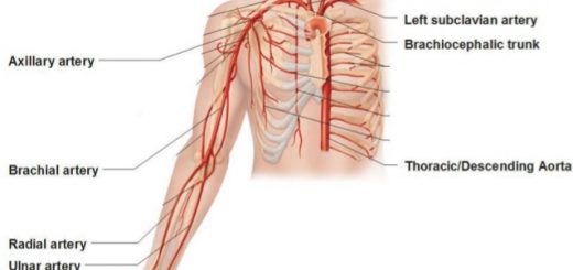
Blood vessels of forearm & hand, Veins and Lymphatics of the upper limb
Forearm bones, anatomy, function & Skeleton of the hand
Nerves of forearm and hand, Nerve injuries types & causes
Flexors of forearm, Forearm muscles, structure, function & anatomy
Arm structure, compartments, muscles, anatomy & Cubital Fossa contents
Bones of upper limb structure, function, types & anatomy
Bone (Osseous Tissue) types, structure, function & importance