Lysis of blood clot, Factors that prevent clot extension and Role of platelets in hemostasis
Platelets share in all steps of hemostasis. Vitamin K is a necessary cofactor for the enzyme that catalyzes one reaction in the synthesis of “vitamin K-dependent” coagulation factors in the liver. Once a blood clot is formed, it can follow two separate courses: either it is invaded by fibroblasts that subsequently from connective tissue, or it can undergo lysis (i.e. dissolve).
Lysis of blood clot (fibrinolysis)
Plasma proteins contain an euglobulin called plasminogen (profibrinolysin) which when activated, is converted to plasmin (fibrinolysin). Plasmin is a proteolytic enzyme that digests fibrin threads and other substances in the clot such as prothrombin. Thus plasma can cause complete lysis (dissolution) of the blood clot.
When a clot is formed, a plasminogen is trapped in the clot along with other plasma proteins. This will not cause lysis of the clot until activated. The injured tissues and endothelium slowly release a powerful activator called tissue plasminogen activator (t-PA) that a few days later, after the clot has stopped the bleeding, converts plasminogen to plasmin, which removes the remaining unnecessary blood clot.
Many small blood vessels in which blood flow has been blocked by clots are reopened by this mechanism. Plasminogen activators are used in the treatment of cardiac and cerebral thrombosis to re-establish blood flow to vital organs.
Factors that prevent clot extension
Normally, endogenous anticoagulant mechanisms prevent clots from becoming unnecessary large by inhibition of activated clotting factors. This is accomplished by several systems that depend on an intact endothelial cell surface.
Tissue factor pathway inhibitor (TFPI): TFPI is a plasma protein that binds to the complex [tissue factor + Factor VIIa + Ca+2] in the extrinsic pathway and blocks the activity of Factor VIIa. TFPI is linked to the endothelial membrane, where it maintains a natural anti-thrombotic surface.
Antithrombin III is a plasma protein that inhibits thrombin and many other enzymes in the coagulation pathway. Heparin, the natural anticoagulant contained in basophils and mast cell granules, is also found on the surface of endothelial cells. It inhibits thrombin by enhancing the activity of antithrombin III.
Protein C is another protein produced in the liver. It inhibits the activity of factors V & VIII. Healthy endothelial cells produce thrombomodulin, which binds to thrombin forming thrombomodulin- thrombin complex that activates protein C.
Paracrine factors: Endothelial cells generate prostacyclin which promotes vasodilation and inhibits platelet activation. nitric oxide which inhibits platelet adhesion and aggregation (released by the endothelium in response to thrombin).
What prevents blood clotting in non-injured vessels?
- Clotting factors circulate in an inactive form.
- The glycoprotein coat of platelets repulses the normal endothelium.
- The healthy endothelium ensures that blood does not come in contact with negatively charged surfaces (e.g. collagen) that activate the intrinsic pathway.
- Blood does not normally come in contact with tissue factors that activate the extrinsic pathway.
- The rapid flow of blood does not allow any activated clotting factors to accumulate.
- There are natural anticoagulants in plasma, including anti-thrombin III and heparin.
- Plasma contains plasminogen, which tissue plasminogen activator (t-PA) can convert to plasma, a protease that digests fibrin and thereby lyses blood clots.
Role of vitamin K in coagulation
Vitamin K is a necessary cofactor for the enzyme that catalyzes one reaction in the synthesis of “vitamin K-dependent” coagulation factors in the liver. They are factor II (prothrombin), VII, IX, and X. Vitamin K deficiency impairs the formation of these factors, resulting in bleeding tendency.
Role of platelets in hemostasis
They share in all steps of hemostasis (vascular spasm, platelet plug formation, coagulation, and clot retraction).
Hemostasis, vascular spasm, clot retardation, Formation of platelet plug & blood clot
Platelets definition, function, structure, normal range & Thrombopoiesis
Blood transfusion causes, Haemolytic transfusion reaction & Acute renal failure
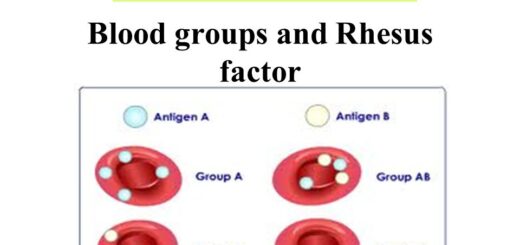
Blood groups, Rh blood groups, Erythroblastosis fetalis & Importance of Rh factor
Symptoms of hemophilia, Disorders of hemostasis, Thromboembolic & Bleeding