Hemostasis, vascular spasm, clot retardation, Formation of platelet plug and blood clot
Hemostasis means the prevention of blood loss or the stoppage of bleeding. When the vessel is ruptured, hemostasis is achieved by several mechanisms. Platelets are necessary for clot retraction to occur. Resting platelets are smooth and discoid shaped. Activated platelets are swollen, sticky with finger-like processes, and secretory. Activation of the platelet contractile proteins can cause a strong concentration of the platelets attached to the fibrin.
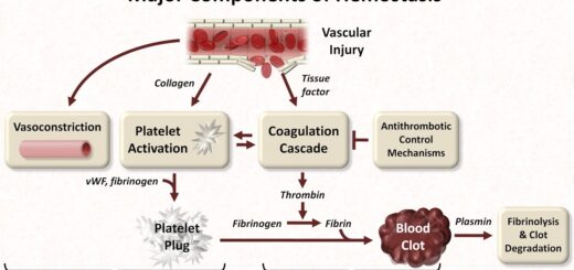
Mechanisms of hemostasis
The steps of hemostasis are vascular spasm, the formation of the platelet plug, the formation of the blood clot (coagulation), and clot retardation.
vascular spasm
Immediately after a blood vessel is cut, the vessel wall contracts, this reduces blood flow from the ruptured vessel. The contraction results from:
- Nervous reflexes initiated by pain.
- Local myogenic contraction of the blood vessels.
- Humoral factors from the traumatized tissues and platelets. Platelets mediate much of the vasoconstriction by releasing the vasoconstrictor substances, thromboxane A2 & serotonin, Also, thrombin that is generated in the coagulation cascade, triggers the endothelium to release the powerful vasoconstrictor, endothelin-1.
This local vascular spasm can last for many minutes or even hours during which the process of platelet plugging and blood coagulation can take place.
Formation of the platelet plug
When the receptors on the platelet membrane come in contact with damaged endothelium or collagen fibers in the vascular wall, the platelets immediately change their shape. They become sticky and develop finger-like processes so that they stick to the collagen fibers, a process called platelet adhesion. This is followed by platelet activation. Activated platelets release the contents of their dense granules, including ADP, thromboxane A2, and serotonin.
These 3 agents in turn act on nearby platelets to activate them, causing them to adhere to the originally activated platelets, resulting in platelet aggregation. Thus, a platelet plug is formed, which is usually successful in stopping blood loss if the vessel opening is small. If there is a large hole, a blood clot is required.
Formation of the blood clot (coagulation)
The clot begins to develop a few seconds after the vascular injury. Coagulation factors are essential to coagulation. They are mostly plasma proteins (inactive proteolytic enzymes) synthesized by the liver. However, some are not protein (e.g. calcium ions) and some are released by the platelets.
Steps of coagulation
- Formation of prothrombin activators (active factor X, active factor V, Ca+2, and phospholipids), This is done by either the intrinsic or the extrinsic pathways.
- Conversion of prothrombin into thrombin (by prothrombin activators).
- Conversion of soluble fibrinogen into insoluble fibrin threads (by thrombin).
Steps 2 and 3 are called the common pathway.
Formation of prothrombin activators
In the body, two different pathways start coagulation:
- The intrinsic pathway becomes activated when blood comes into contact with a rough surface (contact activation). In the lab, this occurs when putting blood into a glass test tube.
- The extrinsic pathway is activated when blood comes in contact with the material from damaged tissues (tissue factor activation). i.e. the stimulus is trauma to the blood vessel wall or surrounding tissues.
In both cases, a chain reaction is triggered in which inactive coagulation factors in the circulation, are sequentially activated. The extrinsic and intrinsic pathways converge on a that ends by converting the soluble plasma protein, fibrinogen (coagulation factor I), to insoluble fibrin, the main constituent of the clot.
The intrinsic pathway (contact activation)
It starts by activation of factor XII by contact of blood with collagen fibers under the injured endothelium. This activation is catalyzed by high-molecular-weight kininogen and prekallikrein. Active factor XII then activates factor XI and active factor XI activates factor IX, Activated factor IX forms a complex with active factor VIII, which activates factor X, Phospholipids from platelets (PL) and Ca+2 are necessary for full activation of factor X.
It is believed that at first, small amounts of factor X are activated without the help of factor VIII. Then, when factor Xa activates prothrombin to thrombin, thrombin activates factor VIII. This results in the feedback activation of the coagulation cascade, with the formation of more thrombin and further activation of factor VIII.
The extrinsic pathway of blood clotting (tissue factor activation)
It is initiated by factors outside the vascular system. Nonvascular cells express the membrane protein tissue factor (tissue thromboplastin, or factor III). Tissue factor acts as a receptor for factor VII, which is a plasma protein. When injury to the endothelium allows factor VII to come in contact with tissue factor, tissue factor activates factor VII. Tissue factor, factor VIIa, and Ca+2 from a complex that proteolytically activates factor X to factor Xa. Factor Xa, which arises by both the intrinsic and extrinsic pathways, proceeds along the common pathway.
The final common pathway
Prothrombin activators (Factor Xa from the extrinsic or intrinsic pathway, factor Va, Ca+2, and phospholipids) convert prothrombin (factor II, one of the plasma proteins) into thrombin. Thrombin (proteolytic enzyme) catalyzes the conversion of soluble fibrinogen (factor I) to insoluble fibrin threads.
Thrombin first converts fibrinogen to fibrin monomers, then many fibrin monomers polymerize into fibrin threads that form the clot. This reaction requires Ca++. Thrombin also activates factor XIII (fibrin-stabilizing factor), which mediates the covalent crosslinking of the fibrin polymers to form a mesh called stable fibrin that is even less soluble than fibrin polymers.
There is a cross-talk between the intrinsic and extrinsic pathways. After blood vessels rupture, clotting is initiated by both pathways simultaneously. However, the extrinsic pathway is very fast, particularly if a large amount of tissue factor is available, while the intrinsic one is much slower. In addition, both pathways augment each other. For example, the [tissue factor + Factor VIIa + Ca+2] complex of the extrinsic pathway, activates factors IX and XI of the intrinsic pathway.
The role of calcium ions in the intrinsic & extrinsic pathways
Except for the first two steps in the intrinsic pathway, calcium ions are required for the promotion or acceleration of all blood-clotting reactions. Therefore, in the absence of calcium ions, blood clotting does not occur by either pathway. In the body, the calcium ion concentration rarely falls low enough to impair blood clotting. But, reducing the calcium ion concentration in a blood sample prevents it from clotting.
Clot retraction
After 20-60 minutes of clot formation, the clot begins to contract and expresses most of the fluid from the clot. The fluid expressed is called serum, which is similar to plasma but without all its fibrinogen and most of the other clotting factors. Thus serum cannot clot because of the lack of these factors. Platelets are necessary for clot retraction to occur.
Role of platelets in clot retraction
They become attached to the fibrin threads in a way that they physically bind different threads together. Platelets entrapped in the clot secrete some of the fibrin stabilizing factor (factor XIII), which forms more cross-linking bonds between the fibrin threads. Activation of the platelet contractile proteins can cause a strong concentration of the platelets attached to the fibrin.
Platelets definition, function, structure, normal range & Thrombopoiesis
Blood transfusion causes, Haemolytic transfusion reaction & Acute renal failure
Blood groups, Rh blood groups, Erythroblastosis fetalis & Importance of Rh factor
Functions and sources of Folic acid, Cobalamin (vitamin B12) & Vitamin K
Factors that help iron absorption, Daily iron requirements & Abnormal iron levels
Lysis of blood clot, Factors that prevent clot extension & Role of platelets in hemostasis