Hemoglobin structure, review and Types of normal hemoglobin
Hemoglobin consists of Heme (It is an iron-porphyrin compound composed of Porphyrins and iron), and Globin (protein part or apoprotein), The transport of O2 is based on a physical interaction between molecular O2 and iron of heme to provide reversible association. Types of normal hemoglobin are Adult hemoglobin, Glycosylated hemoglobin (Hb A1c), and Fetal hemoglobin.
Hemoglobin structure
Heme is an iron-porphyrin compound composed of Porphyrins and iron, Porphyrins are cyclic compounds derived from the porphin nucleus made of 4 pyrrole rings linked by 4 methenyl bridges (-CH=) labelled α, β, γ, and δ. The porphyrins found in nature are compounds in which side chains are substituted for the hydrogen atoms in the porphin nucleus. These are different types of porphyrins. Only types I and III occur in nature:
Type I isomer: The substituent groups attached to the 4 pyrrole rings are symmetrically arranged such as (AP.AP.AP.AP). Type III isomer: The substituent groups attached to the 4th pyrrole ring are arranged in the reverse order, e.g. (AP, AP, AP, PA). The biologically important porphyrin in heme and cytochrome are type III isomers.
Iron is present in the ferrous state (Fe++) and is linked to 4 nitrogen atoms of 4 pyrrole rings. Also, there are 2 additional bonds called 5th and 6th coordination bonds. These two bonds are located on each side of the heme plane (perpendicular to the heme plane).
The 5th position is linked to the nitrogen atom of the imidazole ring of proximal histidine. while the 6th position is bound to oxygen in HbO2 and empty in deoxyhemoglobin (Hb) (unoccupied). The 4 pyrrole rings are attached to side chains called methyl, Vinyl, methyl, Vinyl, methyl, propionyl, propionyl, methyl. (M, V, M, V, M, P, P, M).. The transport of O2 is based on a physical interaction between molecular O2 and iron of heme to provide reversible association.
Globin (protein part or apoprotein) is a simple protein (histone) which is characterized by its high content of histidine and lysine, Globin is composed of four polypeptide chains 2α & 2β chains, The α-chain contains 141 amino acids & β-chain contains 146 amino acids.
Each β-polypeptide chain is folded into 8 right-handed termed A-H starting from NH2-terminal, while α-subunit is folded into 7α-helices. The ratio of heme to globin is 4: 1. So, each heme moiety is linked to one peptide chain.
The myoglobin-hemoglobin family of proteins has produced a way in which Fe++ can be bounded to the proteins so as to produce an O2 binding site. Hemoglobin protects the O2 binding Fe++ from irreversible oxidation by providing environments in which the first step of an oxidation reaction ( the binding of oxygen) is permitted, but the final step (oxidation) is blocked.
Types of normal hemoglobin
Adult hemoglobin. There are 2 types of HbA1 and HbA2. Major adult hemoglobin: Hb A1 (α2 β2) contains 2 alpha chains and 2 beta chains. This hemoglobin A1 constitutes 95-97% of the total hemoglobin. Minor adult hemoglobin: Hb A2 (α2 δ2) contains 2 α-chains and 2 δ-chains. Hb A2 forms about 2-4% of total hemoglobin. In the δ-chains, there is more than one amino acid different than those in β-chains. e.g. arginine residue at the position 16 instead of glycine which is normally present in the beta chain.
Glycosylated hemoglobin (Hb A1c) is a modified form of hemoglobin similar to hemoglobin A1 but it contains glucose linked to ε amino group present on lysyl residues and at the NH2-terminal ends. The reaction is non-enzymatic and its rate depends on the concentration of glucose.
It is present in normal value 5% of the total hemoglobin. This percentage is increased in prediabetic and diabetic patients up to 8-14 %. Thus, glycohemoglobin gives an idea about the blood glucose level during the last three months and is useful in the assessment of diabetic control.
Fetal hemoglobin = HbF (α2γ2) is present normally in newborn and early fetal life. At the age of 7 months, 90 % of fetal hemoglobin is replaced by adult hemoglobin (HbA1). It contains 2 alpha chains and 2 gamma chains. In the gamma chain, there is more than one amino acid different from those in β-chain e.g. His21 residue is ser21. HbF has a great affinity for O2 under physiological conditions because γ-chains do not bind 2,3 BPG well. BPG is responsible for lowering the O2 affinity of Hb and allowing Hb to release O2 at the typical PO2 of tissues.
Blood constituents & Physical properties, Sources & functions of plasma proteins
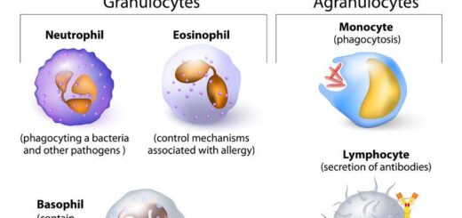
Red blood cells (Erythrocytes) structure & function, Myeloid tissue & Bone marrow
Erythropoiesis, Hemopoiesis, Hemoglobin & Roles of red cells in oxygen transport
Abnormal types of hemoglobin, Sickling of RBCs, Types & causes of methemoglobinemia
Heme biosynthesis & Disorder, Types of porphyrias, Fate of RBs & Catabolism of Hemoglobin