Cerebellum function, structure, anatomy, location and blood supply
The Cerebellum consists of two large hemispheres, which are united by a midline vermis. It presents an anterior notch occupied by the brainstem and a posterior notch occupied by flax cerebelli, It is the largest part of the hindbrain. It is roughly spherical in shape, but its greatest diameter is transverse. In adults, the weight ratio of the cerebellum to the cerebrum is 1:10; in infants, this ratio is 1:20.
Features of the Cerebellum
It occupies the posterior cranial fossa, where it is covered by the tentorium cerebelli. It lies dorsal to the pons and medulla; being separated from them by the cavity of the 4th ventricle. Three pairs of cerebellar peduncles connect the cerebellum with the three segments of the brainstem.
The cerebellum presents two surfaces; superior and inferior. Each surface presents a vermis. On the superior surface, the superior vermis is continuous with the 2 cerebellar hemispheres. While on the inferior surface, the 2 cerebellar hemispheres are separated by a groove called vallecula cerebelli, which is occupied by the inferior vermis.
Fissures of the cerebellum
- Horizontal fissure: separates the superior from the inferior surface.
- The primary fissure separates the anterior lobe from the posterior lobe.
- Posterolateral fissure separates the posterior lobe from the flocculonodular lobe.
- Retrotonsillar fissure: bounding the tonsil.
- Prepyramidal fissure is between the pyramid and the tuber.
- Postpyramidal fissure is between the pyramid and the uvula.
The postpyramidal and the retrotonsillar fissures constitute the secondary fissure or fissure secunda.

Cerebellum
Lobes of the cerebellum
The cerebellum consists of 3 lobes; each one has a central part called vermis and 2 lateral extensions called the cerebellar hemispheres. These lobes are:
- The anterior lobe is in front of the primary fissure.
- The posterior lobe is between the primary fissure and the posterolateral fissure.
- Flocculonodular lobe: consists of nodules and flocculus.
The term corpus cerebelli (body) includes the anterior and posterior lobes.
Internal structure of the cerebellum
The cut section of the cerebellum consists of:
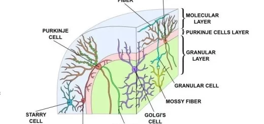
a. Outer cortex of grey matter, the cerebellar cortex.
b. Medullary core of white matter contains 4 pairs of intrinsic nuclei arranged from lateral to medial as follows:
- Dentate nucleus.
- Embolifarm nucleus.
- Globose nucleus. The 2 middle nuclei (emboliform and globose) are called interposed nuclei or nuclei Interpositus.
- Fastigial nucleus. It is commonly called the roof nucleus as it lies in the roof of the fourth ventricle.
The outer cerebellar cortex is organized into groups of folia, termed lobules, that are separated from one another by fissures. Hence, the name folia rather than gyri is used to describe the convolution. In a section through the vermis, the lobules appear to radiate from the apex of the roof of the 4th ventricle. Anatomists recognize ten lobules whose nomenclature is used largely by specialists studying the cerebellum.
Two fissures are particularly deep and divide the various lobules into three lobes: The primary fissure separates the anterior lobe from the posterior lobe, The flocculo-nodular lobe is separated from the posterior lobe by the posterolateral fissure, Beneath the cerebellar cortex is the white matter, which contains axons coursing to and from the cortex, The branching pattern of the white matter of the cerebellum Inspired early anatomists to refer to it as the arbor vitae.
The white matter of the cerebellum consists of:
- The intrinsic fibers do not leave the cerebellum but connect different regions of the organ, Some interconnect folia of the cerebellar cortex and vermis on the same side; others connect the two cerebellar hemispheres together.
- Input (afferent) fibers to the cerebellar cortex: they enter the cerebellum mainly through the inferior and middle cerebellar peduncles.
- Output (efferent) from the cerebellar cortex; these are the axons of the Purkinje cells: The great majorities of the Purkinje cell axons pass to and synapse with the neurons of the deep cerebellar nuclei. A few Purkinje cell axons in the flocculonodular lobe and in parts of the vermis/bypass the cerebellar nuclei and leave the cerebellum without synapsing to reach the vestibular nuclei, The fibers from the dentate, emboliform, and globose nuclei leave the cerebellum through the superior cerebellar peduncle. The fibers from the fastigial nucleus leave through the inferior cerebellar peduncle.
The cerebellar peduncles
These are:
I. Superior cerebellar peduncle: forms a connection between the midbrain and cerebellum, through which pass:
a. Afferent fibers of:
- Ventral spinocerebellar tract.
- Tectocerebellar fibers.
b. Efferent fibers: most of them cross the midline and divide into:
1. Ascending fibers:
- Cerebello-rubral.
- Cerebello-thalamic.
2. Descending fibers:
- Cerebello-olivary.
- Cerebello-reticular.
II. Middle cerebellar peduncle: forms a connection between the pons and cerebellum, through which pass: Afferent fibers: cortico-ponto-cerebellar.
III. Inferior cerebellar peduncle: forms a connection between the medulla oblongata and cerebellum, through which pass:
a. Afferent fibers of:
- Dorsal spino-cerebellar tract.
- Cuneo-cerebellar tract.
- Olivo-cerebellar: from the inferior olivary nucleus of the opposite side.
- Vestibulo-cerebellar: from the vestibular nucleus of the same side.
- Reticulo-cerebellar: The projection of the reticular formation to the cerebellum is bilateral but mainly ipsilateral.
b. Efferent fibers of:
- Cerebello-vestibular.
- Cerebello-reticular.
Blood supply of the cerebellum
A. Arterial supply:
- Superior cerebellar artery: arising from the basilar artery.
- Anterior inferior cerebellar artery: arising from the basilar artery, inferior surface. It supplies part of the inferior surface.
- Posterior inferior cerebellar artery: arising from the vertebral artery. It supplies part of the inferior surface of the cerebellum, including the cerebellar tonsil; it also supplies the dorsolateral surface of the medulla.
B. Venous drainage: Cerebellar veins have a course similar to that of the arteries. They drain into the surrounding venous dural sinuses.
FAQ about the cerebellum
What is the cerebellum?
The cerebellum is a major part of the brain located at the back of the skull. It plays a key role in coordinating movement, balance, posture, and fine motor control. Although it does not initiate movement, it fine-tunes motor activity to make actions smooth and accurate.
Where is the cerebellum located?
The cerebellum sits in the posterior cranial fossa, beneath the occipital lobes of the cerebrum and behind the brainstem. It is separated from the cerebrum by a fold of dura mater called the tentorium cerebelli.
What are the main functions of the cerebellum?
The cerebellum is responsible for:
- Coordinating voluntary movements.
- Maintaining balance and posture.
- Regulating muscle tone.
- Timing and precision of movements.
- Motor learning (improving skills with practice).
- Contributing to eye movements and speech coordination.
What is the structure of the cerebellum?
Structurally, the cerebellum is divided into:
- Two hemispheres (right and left).
- Vermis (the midline part connecting the hemispheres).
- Three lobes: anterior lobe, posterior lobe, and flocculonodular lobe.
It has a highly folded outer layer called the cerebellar cortex and inner white matter that carries signals.
What is the anatomy of the cerebellum?
Anatomically, the cerebellum consists of:
- Cerebellar cortex (gray matter): processes information.
- White matter (arbor vitae): conducts signals within the cerebellum.
- Deep cerebellar nuclei: relay processed signals to other brain regions.
- Cerebellar peduncles (superior, middle, inferior): connect the cerebellum to the brainstem.
How does the cerebellum connect to the rest of the brain?
The cerebellum connects to the brainstem through three paired fiber bundles called cerebellar peduncles. These pathways carry sensory input to the cerebellum and motor output from the cerebellum to the motor areas of the brain and spinal cord.
What is the blood supply of the cerebellum?
The cerebellum receives blood from three main paired arteries:
- Superior cerebellar artery (SCA).
- Anterior inferior cerebellar artery (AICA).
- Posterior inferior cerebellar artery (PICA).
These arteries arise from the vertebrobasilar system and supply different regions of the cerebellum and nearby brainstem structures.
What happens if the cerebellum is damaged?
Damage to the cerebellum can cause:
- Poor coordination (ataxia).
- Unsteady gait and balance problems.
- Intention tremor (tremor during movement).
- Slurred or uncoordinated speech (dysarthria).
- Difficulty with fine motor tasks (e.g., buttoning clothes).
Is the cerebellum involved in thinking or emotions?
While the cerebellum is primarily involved in motor control, research suggests it also contributes to cognitive functions such as attention, timing, and aspects of language, as well as modulation of emotional responses.
How is cerebellar disease diagnosed?
Diagnosis typically involves neurological examination (coordination and balance tests) and imaging studies such as MRI to assess structural changes, strokes, tumors, or degenerative conditions affecting the cerebellum.
Can cerebellar function improve after injury?
Yes, improvement is possible through rehabilitation. Physical therapy, balance training, and occupational therapy can help the brain adapt (neuroplasticity) and improve coordination and daily functioning over time.
You can subscribe to Science Online on YouTube from this link: Science Online
Physiology and functions of Cerebellum, Cerebellar lesions, Motor and Non-motor functions
Cerebellar cortex structure, function, location, layers and fibers
White matter of the brain function, structure and Blood supply of the internal capsule
Anatomy of basal nuclei (basal ganglia) & Disorders of basal ganglia motor circuits
Function and Physiology of Thalamus, Hypothalamus and Limbic system
Diencephalon function, Thalamus, Metathalamus, Hypothalamus, Epithalamus & Subthalamus
Upper and lower motor neurons lesion, Stages of complete spinal cord transection