Histological structure of salivary glands, Parotids, Sublingual and Submandibular glands
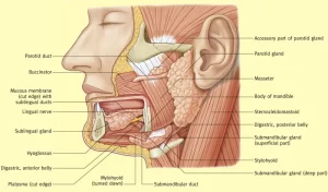
The salivary glands produce saliva through a system of ducts, you have three paired major salivary glands (parotid, submandibular, and sublingual), as well as hundreds of minor salivary glands, In humans, 1200 to 1500 ml of saliva are produced every day, The secretion of saliva (salivation) is mediated by parasympathetic stimulation; acetylcholine is the active neurotransmitter and binds to muscarinic receptors in the glands, leading to increased salivation.
Histological structure of salivary glands
The mouth cavity is continuously lubricated and washed out by saliva. It is the secretory product of a group of salivary glands that open into the oral cavity. Saliva aids in digestion, It keeps your mouth moist, and supports healthy teeth. Salivary glands are generally categorized under two groups:
A. The main (major) salivary glands
These include three pairs of glands; the parotids, the sublingual, and the submandibular glands. They are large compound tubuloalveolar (tubuloacinar) glands, which pour their products via a branched system of ducts. Their secretion is regulated by nervous reflexes.
B. The accessory (minor) salivary glands
These include unpaired glands; the buccal, labial, lingual, and palatine glands, They are small, simple tubuloalveolar glands scattered in the oral mucosa, They secrete continuously to moisten and lubricate the oral cavity.
Histological organization of the main salivary glands
The salivary gland is constructed of stroma and parenchyma.
The stroma
Connective tissue capsule that covers the gland, separating it from the surrounding structures and conveying its vascular and nervous supply. Connective tissue septa divide the gland into lobes, by interlobar septa and smaller lobules, by interlobular septa. The intralobular connective tissue is formed of a network of reticular fibers that surround the secretory acini and their ducts.
The parenchyma
A. The secretory acini: the acinus = group of secretory cells surrounding a lumen and resting on an abasement membrane. The cells are separated from the basement membrane by the myoepithelial cells.
Myoepithelial cells (basket cells)
- They are stellate epithelial cells with many processes that surround the acini, like a basket.
- They are also located along the proximal portions of the ducts.
- They can contract, by their actin and myosin to squeeze the secretion from the acini into the lumen of their ducts.
There are three types of acini found in the salivary glands:
1. Mucous acini
They secrete mucus (viscid glycoprotein) secretion. They are present in the sublingual submandibular, buccal, labial, and palatine glands.
Histological features of a mucous acinus
- The mucous acinus appears large, pale, and oval with a wide lumen.
- Each acinus is lined by cuboidal cells with flattened, basal nuclei and pale vacuolated cytoplasm as a result of the dissolution of the mucinogen granules during the processing of the H&E section.
- Each mucous acinus is surrounded by numerous basket cells to help the drainage of the thick mucus into the ducts.
2. Serous acini
They produce watery secretion rich in salts and digestive enzymes. They are present in the parotid, submandibular, and Von Ebner’s glands.
Histological features of a serous acinus
- The serous acinus appears small, deeply-stained, and rounded with a narrow lumen.
- Each acinus is lined by pyramidal cells with rounded nuclei situated nearer to the base. The serous secretory cells have and basal basophilic cytoplasm (rich in rER) with apical eosinophilic secretory (zymogen) granules.
- The secretory acinus is surrounded by a few basket cells as their watery secretion flows easily through the ducts.
Mixed (seromucous) acini
They are present in the submandibular and a few in the sublingual glands. In routine histological preparation, the mixed acinus appears as a mucous with a cap of serous cells (serous demilunes). Recent microscopic studies indicated that each acinus is formed of mucous cells with some serous cells interspersed among them. Both cells pour their secretion into the lumen of the acinus.
B. The duct system:
1. The intralobular ducts formed of 2 parts:
- The intercalated ducts: they drain the secretory acinus and are lined by low cuboidal cells.
- The secretory striated ducts: the intercalated ducts unite together to pour into larger secretory striated ducts. They appear with a wide lumen lined by cuboidal cells with central rounded nuclei and basal eosinophilic striated cytoplasm due to the presence of numerous mitochondria lying in basally-infolded cell membrane The secretory striated ducts modify the composition of saliva by active ion pump making the saliva hypotonic solution.
2. The interlobular ducts: the intralobular ducts unite to form larger interlobular ducts lying in the interlobular connective tissue septa. They are lined by columnar epithelium.
3. The interlobar ducts: larger ducts in the interlobar septa, They are lined by pseudostratified columnar epithelium.
4. The main duct: large duct with a wide lumen. It is lined at its beginning by stratified columnar epithelium which gradually becomes non-keratinized stratified squamous epithelium similar to that lining oral cavity where it opens.
Histological features of the parotid gland
- Capsule & septa: thick, well-defined lobulation.
- Fat cells: large number within the septa and between the acini.
- Secretory acini & myoepithelial cells: in human, 100% serous acini with relatively few myoepithelial cells.
- The intralobular ducts: long, secretory striated ducts form 50% of the length of the intralobular duct.
Histological features of the submandibular gland
- Capsule & septa: moderately thick.
- Fat cells: less numerous.
- Secretory acini & myoepithelial cells: 80% serous acini, 20% mucous acini with frequent myoepithelial cells.
- The intralobular ducts: moderately-long, secretory striated ducts form 80% of the length of the intralobular duct.
Histological features of the sublingual gland
- Capsule & septa: thin capsule, relatively thick septa. Well-defined lobulation
- Fat cells: no fat cells
- Secretory acini & myoepithelial cells: 99% mucous, 1% serous, represented by the serous demilunes surrounded by numerous myoepithelial cells.
- The intralobular ducts: very short, secretory striated ducts form 99% of the length of the intralobular duct.
You can download Science online application on Google Play from this link: Science online Apps on Google Play
Git function, Types of salivary secretion and Composition of saliva
Norma lateralis features and anatomy, Mandible surfaces, nerves and ligaments
Salivary glands function, shape, lobes, surfaces and Structures within the parotid gland
Mouth Cavity divisions, anatomy, function, muscles, Contents of Soft palate and Hard palate
Tongue function, anatomy and structure, Types of lingual papillae and Types of cells in taste bud


