Norma lateralis features and anatomy, Mandible surfaces, nerves and ligaments
The skull is a shell for the brain and the origins of the central nervous system, it includes all the bones of the head, face, and jaws, there are twenty-eight individual bones, eleven of them are paired, to form a bilaterally symmetrical three-dimensional structure and six of them are single, unique bones, The skull bones are categorized into regional and development bones, the skull is classified by its two main parts: the cranium and the mandible.
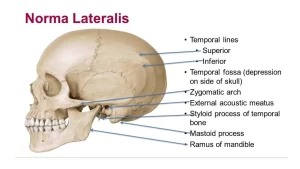
Norma lateralis
Bones forming it:
- Superiorly: Nasal, Frontal and parietal bones.
- Inferiorly: Maxilla, zygomatic, greater wing of sphenoid, squamous, and mastoid parts of the temporal bone.
Features:
The squamous part of the temporal bone articulates in front with the greater wing of the sphenoid and upwards with the parietal bone at the parieto-temporal suture. This suture meets the lambdoid suture posteriorly at a point called Asterion.
Zygomatic Arch:
It is formed by the temporal process of the zygomatic bone and the zygomatic process of the temporal bone.
Temporal line:
The posterior border of the zygomatic bone is sharp and gives the temporal line. this continues upwards then arches back and diverges into superior and inferior temporal lines. The superior temporal line is traceable till the mastoid process. The temporalis fascia is attached to this line. The temporal fossa is enclosed by the inferior temporal line.
The mandible
It is the skeleton of the lower jaw and the only mobile bone of the skull. It is formed of a body and two rami.
The body of the mandible:
It is horse-shoe shaped having two margins (an alveolar margin and a lower border) and two surfaces (outer & inner). The upper border: carries the sockets for 16 teeth. The base of the mandible: continues posteriorly with the posterior border of the ramus. It shows 2 digastric fossae on either side of the midline anteriorly.
The surfaces
A. The outer surface: it has the following features:
- The symphysis menti: a faint median ridge representing the site of fusion between the two halves of the mandible (at one year to 4 years).
- Mental protuberance is a triangular raised area at the lower part of the symphysis menti.
- Mental foramen: it lies opposite the line between the two premolars. It is the anterior end of the mandibular canal. It transmits the mental nerve and vessels.
- The oblique line extends from the anterior border of the ramus to the mental foramen.
B. The inner surface: It shows the following:
- Superior and inferior genial tubercle: present at the lower part of the inner surface, close to the midline.
- Mylohyoid line: It begins below and behind the last molar tooth. Then extends downwards to symphysis menti. It divides the inner surface into two fossae.
- Sublingual fossa: above and in front of the mylohyoid line. It is related to the sublingual salivary gland.
- Submandibular fossa: It lies below and behind the mylohyoid line. It is related to the submandibular salivary gland.
Ramus of the mandible
It projects upwards on either side from the posterior part of the body. It has 4 borders, 2 surfaces & 2 processes.
Borders:
- Upper border: it forms the mandibular notch.
- Lower border: It meets the posterior border at the angle of the mandible.
- Posterior border: It extends from the angle to the back of the condylar process.
- Anterior border: It is continuous above with the anterior border of the coracoid process and below with the oblique line of the body.
Surfaces:
- Outer (lateral) surface.
- Inner (medial) surface.
a. Mandibular foramen: the center of ramus and leads to the mandibular canal. It transmits the inferior alveolar nerve and vessels. The margin of the foramen is irregular, it presents a prominence called lingula.
b. Mylohyoid groove: It starts below the mandibular foramen and passes downwards and forwards to end below the posterior end of the mylohyoid line of the body.
c. Condylar process: It is an upward flattened projection from the posterior part of the ramus. It consists of:
- Head of the mandible: it articulates the mandibular fossa of the skull forming tempro-mandibular joint.
- The neck of the mandible: It is the constriction below the head. Its antero-medial aspect shows a depression called pterygoid fossa or fovea.
Nerves related to the mandible
1. Nerves passing in foramina:
- Inferior alveolar nerve: it passes through the mandibular foramen and canal where it divides into the incisive nerve (supplying the teeth) and the mental nerve
- The mental nerve: passes through the mental foramen.
2. Nerves related to grooves on the mandible:
- Nerve to mylohyoid nerve in the mylohyoid groove.
- The lingual nerve in a groove at the medial aspect of the last molar tooth socket.
3. Nerves related to the neck of the mandible:
- Nerve to masseter: as it passes through the mandibular notch.
- Auriculotemporal nerve: It passes medial to the neck of the mandible.
Ligaments attached to the mandible
- Tempero-mandibular ligament: It extends from the lower border of the zygomatic arch to the lateral surface of the neck of the mandible.
- Stylo-mandibular ligament: From the styloid process to the posterior border of the angle of the mandible.
- Spheno-mandibular Ligament: From the spine of the sphenoid bone to the lingula of the mandible.
- Pterygomandibular ligament: From the pterygoid hamulus to the posterior end of the mylohyoid line of the mandible.
You can download Science online application on Google Play from this link: Science online Apps on Google Play
Histological structure of salivary glands, Parotids, Sublingual and Submandibular glands
Mouth Cavity divisions, anatomy, function, muscles, Contents of Soft palate and Hard palate
Tongue function, anatomy & structure, Types of lingual papillae and Types of cells in taste bud
Salivary glands function, shape, lobes, surfaces and Structures within the parotid gland


