Mouth Cavity divisions, anatomy, function, muscles, Contents of Soft palate and Hard palate
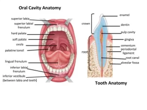
The oral cavity, or the mouth or buccal cavity serves as the first portion of the digestive system, It consists of the lips, tongue, palate, and teeth, The mouth is important to several bodily functions, such as breathing, speaking, chewing, drinking, swallowing, talking, tasting, digesting food and drinks. This oval-shaped opening in your skull starts at your lips and ends at your throat, the mouth allows air and nutrients to enter your body, and it helps you speak.
Divisions of the mouth cavity
The mouth cavity is divided into 2 main parts:
- Vestibule of the mouth.
- Mouth cavity proper.
Vestibule of mouth
It is a slit-like space bound externally by the lips and cheeks, internally by gums and teeth. The vestibule opens into the outside through the oral fissure. When the jaws are closed, the vestibule is connected to the mouth cavity proper through an interval behind the last molar teeth on each side.
Mouth cavity proper
It is bounded by:
- Anterior and laterally: the gums and teeth.
- Posteriorly: it communicates with the pharynx through the isthmus of fauces. This isthmus is bounded by the palatoglossus arch on each side.
- Roof: hard and soft palate.
- Floor: mylohyoid muscle.
It is occupied mainly by the tongue. A median fold of mucosa connects the under the surface of the tongue to the floor of the mouth; this is called the frenulum linguae. The floor presents a small ridge on which contains the sublingual papillae on each side of the frenulum. This ridge is called the sublingual fold. Lateral to the frenulum, there is the deep lingual vein and lateral to it are the fimbriated folds (one on each side).
Muscles of the floor of the mouth
1- Digastric muscle
Origin and insertion: This muscle has a posterior belly, an intermediate tendon, and an anterior belly. The posterior belly arises from the mastoid notch and ends in the intermediate tendon. The intermediate tendon is held in position by a loop of deep fascia, which binds the tendon down to the junction of the body and greater cornu of the hyoid bone. The anterior belly is attached to the digastric fossa near the median plane.
Nerve supply: The posterior belly is supplied by the facial nerve and the anterior belly by the nerve to the mylohyoid (mandibular nerve).
Action: It depresses the mandible or elevates the hyoid bone during deglutition.
1. Stylohyoid muscle: supplied by the facial nerve.
2. Mylohyoid muscle:
- Origin: It arises from the whole length of the mylohyoid line of the mandible.
- Insertion: The posterior fibers are inserted into the body of the hyoid bone; the anterior fibers are inserted into a fibrous raphe in the midline.
- Nerve supply is supplied by the mylohyoid branch of the inferior alveolar nerve.
- Action: When the mandible is fixed, they elevate the floor of the mouth and hyoid bone during the first stage of swallowing, When the hyoid bone is fixed, it assists in the depression of the mandible and the opening of the mouth.
3. Geniohyoid muscle:
- Origin: It takes origin from the inferior genial tubercle of the mandible.
- Insertion. It is inserted into the anterior surface of the body of the hyoid bone.
- Nerve supply: C1 component of the hypoglossal nerve.
- Action: Elevates the hyoid bone or depresses the mandible.
The palate
The palate forms the roof of the mouth. It is divided into:
I. Hard palate
It is the bony anterior two-thirds, It is composed of the palatine processes of the maxillae and the horizontal plates of the palatine bones, It is bounded by the alveolar arches and is continuous posteriorly with the soft palate, It forms the floor of the nasal cavity, It is covered with a mucous membrane with an inferior median raphe and bilateral corrugations on both sides.
II. Soft palate
It extends from the posterior border of the hard palate, It is covered with a mucous membrane, It ends posteriorly with the uvula, and is continuous on both sides with the lateral wall of the pharynx.
Contents of the soft palate:
- Palatine aponeurosis.
- Muscles.
- Nerves.
- Vessels.
- Lymphoid tissue.
Palatine apponeurosis
It is a fibrous sheet attached to the posterior border of the hard palate, It is the expanded tendon of the tensor palati muscle, It splits to enclose the musculus uvula muscle, and it gives attachment to all palatine muscles.
Muscles of the soft palate
1- Tensor palate muscle:
- Nerve supply: Trunk of the mandibular nerve.
- Action: Tenses or tighten the soft palate.
2- Levator palati muscle:
- Nerve supply: Cranial part of the accessory nerve through the pharyngeal plexus.
- Action: Elevation of the soft palate. Both muscles open the auditory tule to equalize the pressure between the middle ear and nasopharynx.
3- Palatoglossus muscle:
- Nerve supply: Cranial part of the accessory nerve through the pharyngeal plexus.
- Action: It depresses the palate, elevates the root of the tongue, and narrows the oropharyngeal isthmus (between the mouth cavity and oropharynx).during deglutition.
4- Palatopharyngeus muscle:
- Nerve supply: Cranial part of the accessory nerve through the pharyngeal plexus.
- Action: Depression of the palate and together with the soft palate, it narrows the nasopharyngeal isthmus during deglutition.
5- Musculus uvulae:
- Nerve supply: Cranial part of the accessory nerve through the pharyngeal plexus.
- Action: Retract and elevate the uvula.
Nerve supply of the palate
A- Sensory nerve supply
Hard palate:
- Greater palatine nerve.
- Nasopalatine nerve
Soft palate:
- 3. Lesser palatine nerve.
- 4. Glossopharyngeal nerve.
Secretomotor fibres to the palatine glands
The facial nerve through its greater petrosal nerve relays in the sphenopalatine ganglion, Postganglionic fibers reach the palatine glands through the lesser palatine nerves.
Taste sensation
Taste fibers reach the inferior surface of the palate through the lesser palatine nerves.
B- Motor nerve supply
All the muscles are supplied by the cranial part of the accessory nerve except the tensor palati muscle which is supplied by the mandibular nerve.
Arterial supply of the palate
- Greater palatine artery (a branch from the maxillary artery).
- Ascending palatine artery (a branch from the facial artery).
- Palatine branch of ascending pharyngeal artery. Venous drainage of the palate: Through pterygoid and pharyngeal plexuses of veins.
Lymphatic drainage of the palate
- The hard palate drains into submandibular lymph nodes.
- The soft palate drains into the upper deep cervical and retropharyngeal lymph nodes.
You can subscribe to Science online on YouTube from this link: Science Online
You can download Science online application on Google Play from this link: Science online Apps on Google play
Git function, Types of salivary secretion & Composition of saliva
Histological structure of salivary glands, Parotids, Sublingual and Submandibular glands
Norma lateralis features & anatomy, Mandible surfaces, nerves & ligaments
Tongue function, anatomy & structure, Types of lingual papillae & Types of cells in taste bud
Salivary glands function, shape, lobes, surfaces and Structures within the parotid gland


