Larynx structure, function, cartilages, muscles, blood supply and vocal folds
The larynx protects the lower airways, It facilitates respiration, It plays a key role in phonation, It directs air into the lungs to breathe, It directs food into the esophagus on its way to the stomach, It can create a sound for the voice, vocal cords can open to allow breathing; close to protect the windpipe when swallowing, and vibrate to give voice.
Larynx
The larynx is the organ of phonation (voice production) in addition to its respiratory function (airway). It is formed of a group of cartilages connected by muscles, ligaments, and joints). The larynx lies below the hyoid bone in the midline of the neck at the level of vertebrae.
Sensory nerves to the larynx are derived from the internal branch of the superior laryngeal nerve (iSLN) and the recurrent laryngeal nerves (RLNs); both are branches of the vagus nerve. The RLN innervates all intrinsic laryngeal muscles except the cricothyroid muscle, which is innervated by the external division of the SLN (eSLN).
Reflexive glottic closure is inhibited by hypothermia, inspiratory phase, increased arterial pCO2, decreased arterial pO2, and positive intrathoracic pressure. Tracheostomy leads to centrally mediated impaired reflexes and decannulation (adductor failure and vocal cord fusion).
Cartilages of the Larynx:
Single cartilages Paired cartilages
Thyroid cartilage Arytenoid cartilage
Cricold cartilage Corniculate cartilage
Epiglottic cartilage Cuneiform cartilage
Inlet of the larynx
Boundaries:
- Anterior: Upper edge of the epiglottis.
- On each side: Aryepiglottic folds.
- Posterior: Mucous fold between the arytenoids.
The cavity of the larynx:
The cavity of the larynx is divided into three compartments by two pairs of folds that extend from before backwards, The upper folds are called vestibular folds (false vocal cords) and the lower folds are called vocal folds (true vocal folds). The compartments are:
- Vestibule of the larynx: It is the area between the inlet and the vestibular folds (upper compartment).
- Sinus (ventricle) of the larynx: It is the area between the vocal fold and the vestibular fold on each side. (middle compartment)
- The infraglottic compartment (lower compartment) it lies below the vocal folds and is continuous below the trachea.
Vocal folds
It is the upper free margin of the cricothyroid ligament. It extends between the angle of the thyroid cartilage and the vocal process of the arytenoid cartilage. Rima glottidis is the narrowest part of the laryngeal cavity between the two vocal cords. Rima vestibuli is the space between the two vestibular folds. The saccule of the larynx is an upward recess deep to the vestibular folds.
Muscles of the Larynx:
I. Muscles acting on the laryngeal inlet:
A: Muscles closing the laryngeal inlet:
- Aryepiglottic muscles
- Transverse arytenoid
- Oblique arytenoids
- Thyro-epiglottic
No muscles produce an opening of the laryngeal inlet, it is only opened by the elastic recoil of the epiglottis.
II. Muscles acting on the vocal curds:
A: Muscles producing abduction of the vocal cords:
The posterior crico-arytenoid is the only abductor to the vocal cords. It extends from the posterior surface of the lamina of the cricoid cartilage to the muscular process of the arytenoid. Actions: Abduction of the vocal cords.
B. Muscles producing adduction of the vocal cord:
- Lateral crico-arytenoid.
- Transverse arytenoid.
- Oblique arytenoids
C. Muscles stretching (tensing) the vocal cords: Cricothyroid muscle.
D. Muscles relaxing the vocal cords:
- Thyroarytenoid muscle:
- Vocalis muscle:
Nerve supply of larynx
A. Motor supply: All intrinsic muscles of the larynx are supplied by the “recurrent laryngeal nerve” except cricothyroid which is supplied by the external laryngeal nerve.
B. Sensory supply:
- Above the vocal cords ……………….. Internal laryngeal nerve (from vagus N).
- Below the vocal cords ……………….. Recurrent laryngeal nerve (from vagus N).
Blood supply of larynx
A. Arterial supply:
- Above the vocal cords: Superior laryngeal artery (a branch of the superior thyroid artery).
- Below the vocal cords: Inferior laryngeal artery (a branch of inferior thyroid artery).
B. Venous drainage: It drains its venous blood into the corresponding superior and inferior thyroid veins respectively.
Clinical points:
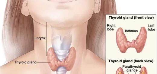
- Injury of the external laryngeal nerve (in thyroidectomy) leads to paralysis of the cricothyroid muscle which results in low pitched voice.
- Injury of the recurrent laryngeal nerve (in thyroidectomy):
- If partial injury, It leads to adduction of the vocal cord. If this occurs bilaterally, it leads to suffocation (obstruction of the airway).
- If complete injury, it leads to the cadaveric position of the vocal cords (midway between abduction and adduction).
The fibers that produce abduction lie in the outer part of the nerve (affected by partial injury), while the fibers that produce adduction lie in the central part of the nerve (affected by complete injury).
Trachea, Major Bronchi, Thoracic cavity structure, function and anatomy
Anatomy of nose, function of para-nasal air sinuses and Sphenopalatine Ganglion branches
Diaphragm anatomy, structure, function, Phrenic nerves and Nerves of the thorax
Respiratory Chain Electron Transport Chain (ETC) components, control and inhibitors
Blood vessels structure, function, layers, characteristics and How blood vessels work