Histological structure of stomach, Fundic glands of Stomach and Gastric musculosa
The stomach store and macerate food, and begin the early phases of food digestion, It is a bag-like muscular organ found on the upper abdomen’s left side, The esophagus delivers food to the stomach through a valve called the esophageal sphincter, It produces acid and enzymes that aid in the digestion of meals.
Histological structure of the stomach
The stomach is the most dilated segment of the digestive tube.
General histological features of the stomach
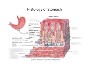
The mucosa and the submucosa of the empty stomach display longitudinal folds, the gastric rugae which disappear in the distended state, Rugae permit expansion of the stomach when it is filled with food and gastric juices. The mucosal surface of the stomach is lined by the surface mucous cells and is invaginated into the lamina propria forming the funnel-shaped gastric pits. The lamina propria contains a large number of branched, tubular gastric glands that open into the bottoms of the gastric pits.
From the histological point of view, the stomach is divided into 3 regions:
- The cardiac region surrounds the entrance of the esophagus and contains the cardiac glands.
- The fundic region is the largest part, including the fundus and the body of the stomach, and contains the fundic glands.
- The pyloric region, is proximal to the pyloric sphincter and contains the pyloric glands.
I- Mucosa of fundic region of the stomach
The Mucosa of fundic region is characterized by short and narrow gastric pits. Fundio mucosa is formed of:
1. The surface mucous cells:
- They are tall columnar cells with pale apical parts, covering the surface of the mucosa and lining the gastric pits in all gastric regions.
- They secrete the visible thick mucus with neutral or slightly alkaline pH. It protects the epithelium from auto-digestion and from abrasion by food.
2- The lamina propria: it is almost occupied by a large number of fundic glands that secrete the main bulk of gastric juice. The glands are surrounded by a little amount of highly vascular, loose connective tissue.
3- Muscularis mucosae: in all gastric regions, it is formed of inner circular and outer longitudinal layers of smooth muscle fibers.
The fundic glands of the stomach
They are closely-packed simple branched tubular glands, extending throughout the full thickness of the mucosa, perpendicular to the muscularis mucosae.
5 types of cells lining the fundic gland
1. The mucous neck cells: they are low columnar and present in the superficial (upper) part of the fundic gland. They secrete soluble mucus to lubricate the gastric content.
2. The oxyntic cells (parietal cells):
- They are present mainly in the middle part of the fundic gland.
- They are pyramidal cells lodged between the basement membrane and the other cells lining the fundic gland.
- They have rounded nuclei with intense eosinophilic cytoplasm rich in mitochondria with no secretory granules.
- They have intracellular canaliculi which are deep invaginations of the apical plasma membrane into the cytoplasm, communicating with the lumen of the gastric gland. The wall of each canaliculus has numerous microvilli projecting into its lumen. An active ion pump occurs across the membranes of these microvilli, which is important for HCI formation.
- The Tubulovesicular system of membranes is present in the cytoplasm adjacent to the canaliculi. In an actively secreting cell, membranes of the tubulovesicular system decrease as they are inserted into the wall of the canaliculi forming numerous microvilli to increase the surface area available for acid production.
- The oxyntic cells secrete HCL into the lumina of the intracellular canaliculi. They also secrete the intrinsic factor, a glycoprotein essential for the absorption of vitamin B12
3. The peptic cells (chief cells, zymogenic cells): they are the most numerous cells in the fundic gland, present mainly in the base of the gland. They are pyramidal cells with a strongly basal basophilic cytoplasm (due to abundant rER) and apical eosinophilic zymogen secretory granules. They secrete pepsin and gastric lipase.
4. Enteroendocrine cells:
- They are located mainly in the base of the fundic gland. They belong to the widely distributed diffuse neuroendocrine system.
- They are small pyramidal cells, either of closed type (lie directly on the basement membrane and do not reach the lumen) or of open type (extend to reach the lumen by their microvilli).
- They contain basal secretory granules close to the adjacent blood capillaries in the lamina propria.
- They are best visualized by immunohistochemical staining for their peptide hormones secretion; such as enterochromaffin cells (EC cells), secreting serotonin, and enterochromaffin-like cells (ECL cells), secreting histamine.
5. Stem cells: they are present mainly in the upper part of the fundic gland. They are low columnar cells usually seen in mitosis. They proliferate and move either superficially to differentiate and replace the surface mucous cells; or deeply where they differentiate into all types of secretory cells lining the gastric glands.
II. Mucosa of cardiac region of the stomach
The cardia is a narrow region of the stomach that surrounds the esophageal orifice. Its mucosa has the following histological features:
- Shallow gastric pits, lined by surface mucous cells.
- The lamina propria contains branched, coiled, tubular cardiac glands
- Cardiac glands are lined mainly by mucus-secreting cells.
III. Mucosa of pyloric region of the stomach
It is characterized by: Wide and deep gastric pits occupying more than half the lengths of the mucosa. The lamina propria contains the branched, coiled, tubular pyloric glands which are lined by mucus-secreting cells and some enteroendocrine cells, particularly G cells secreting gastrin hormone.
Gastric musculosa
It is formed of spirally-oriented smooth muscle fibers, arranged in inner oblique, middle circular, and outer longitudinal layers. The arrangement of muscle layers is related to its role in mixing gastric chyme during the digestive process. In the region of the pylorus, the circular smooth muscle layer is thickened constituting the pyloric sphincter.
Gastric serosa: the entire stomach is invested by serosa.
You can download Science Online application on Google Play from this link: Science online Apps on Google Play
Posterior abdominal wall muscles, layers, blood supply & anatomy
Regulation of stomach emptying, Causes of vomiting & Nervous pathway of reflex vomiting
Physiology & functions of Stomach, Composition of gastric secretion
Stomach parts, function, curvatures, orifices, peritoneal connections & Venous drainage of Stomach
Abdomen muscles, Blood Supply of Anterior Abdominal Wall and Rectus Sheath content
Histological organization of pharynx, Structure and function of esophagus
Mouth Cavity divisions, anatomy, function, muscles, Contents of Soft palate and Hard palate


