Abdomen muscles, Blood Supply of Anterior Abdominal Wall and Rectus Sheath content
The abdomen is commonly called the belly, It is the body space between the thorax (chest) and pelvis, The diaphragm forms the upper surface of the abdomen, The abdominal muscles allow movement and hold organs in place by regulating internal abdominal pressure, and they support the trunk, The deep abdominal muscles, together with muscles in the back, make up your ‘core’ muscles and help keep your body stable and balanced, and protect your spine.
Abdomen
The abdomen is the part of the trunk that lies between the thorax and pelvis. It lies between the diaphragm and the pelvic brim.
Anterior Abdominal Wall
Abdominal Regions
The abdomen is divided into 9 regions by two pairs of planes:
- Vertical Planes: Left and right lateral planes, each extends from mid-clavicular to mid-inguinal point.
- Horizontal Planes:
- Subcostal plane: It lies immediately inferior to the costal margins and passes posteriorly through the body of the L3 vertebra.
- Intertubercular plane: Connects the tubercles of iliac crests and reaches the upper part of the body of L5.

Abdomen muscles
Important landmarks
- Midinguinal point: A point midway between the anterior superior iliac spine and the symphysis pubis.
- Transpyloric plane: At the level of the L1 vertebra and midway between the xiphoid process of sternum and umbilicus.
Abdominal regions are:
- Right hypochondrium.
- Epigastric region.
- Left hypochondrium.
- Right lumbar (flank) region.
- Left lumbar (flank) region
- Umbilical region.
- Right iliac fossa (groin).
- Left iliac fossa (groin).
- Hypogastric region.
Dermatomes of anterior abdominal wall
The dermatome is the skin area supplied by a single segment of the spinal cord. There are “three” anatomical landmarks (levels) for the dermatomal supply of the skin of the anterior abdominal wall, at the xiphoid process T. 7, at the umbilicus T10 and at the suprapubic region T. 12 (above the symphysis pubis).
The fascia of Anterior Abdominal Wall
Superficial Fascia:
- This superficial fascia consists of one layer that contains a variable amount of fat.
- The superficial fascia below the level of the umbilicus can be divided into two layers:
- Superficial fatty layer (Camper’s fascia) containing a variable amount of fat.
- Deep membranous layer (Scarpa’s fascia) containing fibrous tissue and very little amount of fat. It is continuous with colle’s fascia superficial perineal fascia. The superficial vessels and nerves run between these two layers.
Muscles of the anterior abdominal wall
Muscle: External abdomina I oblique.
- Origin: External surface of lower 8 ribs. Its fibers are directed downwards, forwards, and medially.
- Insertion: By an apponeurosis into the xiphoid process, linea alba, pubic crest & tubercle, anterior superior iliac spine (ASIS) & anterior half of iliac crest.
- Action: Muscles of both sides flex the trunk. Muscles of one side bend the trunk to the same side (lateral flexion). Compresses the abdominal contents.
- Innervation: Intercostal nerves 7-11, subcostal, iliohypogastric and ilioinguinal nerves.
Muscle: Internal abdomina I oblique.
- Origin: Thoracolumbar fascia, anterior 2/3 of the iliac crest, lateral 2/3 of the inguinal ligament. Its fibers are directed upwards, forwards, and medially.
- Insertion: Lower 3 or 4 ribs, xiphoid process, linea alba, pubic crest, and pectineal line. Its lower fibers form an arch called a conjoint tendon.
- Action: Muscles of both sides flex the trunk. Muscles of one side bend the trunk to the same side (lateral flexion). Compresses the abdominal contents.
- Innervation: Intercostal nerves 7-11, subcostal, iliohypogastric and ilioinguinal nerves.
Muscle: Transversus abdominis
- Origin: Inner surface of the lower 6 ribs, thoracolumbar fascia, anterior 2/3 of the inner lip of iliac crest, lateral 1/3 of the inguinal ligament.
- Insertion: Linea alba, pubic crest, and pectineal line. Its lower fibers form an arch together with the lower fibers of the internal oblique called the conjoint tendon.
- Action: Muscles of both sides flex the trunk. Muscles of one side bend the trunk to the same side (lateral flexion). Compresses the abdominal contents.
- Innervation: Intercostal nerves 7-11, subcostal, iliohypogastric and ilioinguinal nerves.
Muscle: Rectus abdominis
- Origin: Pubic crest and symphysis pubis.
- Insertion: Xiphoid process, 5th, 6th & 7th costal cartilages.
- Action: Flexes the trunk and raises intra-abdominal pressure.
- Innervation: intercostal nerves 7-11 and subcostal nerve.
Muscle: Pyramidalis
- Origin: Pubic crest anterior to rectus abdominis.
- Insertion: Linea alba.
- Action: Draws the linea alba inferiorly.
- Innervation: Subcostal nerve.
Muscle: Cremasteric Muscle
- Origin: Lower fibers of the internal oblique muscle.
- Insertion: The pubic tubercle, forms a thin network of muscle loops around the spermatic cord and testis (or around the distal portion of the round ligament of the uterus).
- Action: Elevates Testis (not well developed in females).
- Innervation: Genital branch of the genitofemoral nerve.
The inguinal ligament is the infolded lower part of the external oblique aponeurosis. This extends between the ASIS and pubic tubercle. The external spermatic fascia is the external oblique muscle’s contribution to the coverings of the testis and spermatic cord.
Parts of the inguinal ligament
1. Reflected ligament: few fibers pass upwards and medially to the inserted into the linea alba.
2. Lacunar ligament: Its upper border is attached to the medial part of the inguinal ligament while its lower border is attached to the medial part of the pectineal line.
3. Pectineal ligament: Few fibers extend from the lacunar ligament to be attached to the pectineal line.
- Cremasteric muscle and fascia are the internal oblique muscle’s contribute to the coverings of the testis and spermatic cord. Cremasteric reflex may be elicited by stroking the medial thigh (where the femoral branch of the genitofemoral n. distributes cutaneously) (cremaster = a suspender).
- The conjoint tendon is formed of the lower arched fibers of both internal oblique and transversus abdominis muscles.
These arching fibers pass directly from the origin to insertion (run for a short distance).
Function
Contraction of the conjoint tendon tightens it and lowers the roof of the inguinal canal (narrowing it) (shutter mechanism). This narrowing prevents the passage of the intestine through the inguinal canal.
- Transversalis fascia, the deep fascia that covers the inner surface of the transversus abdominis, forms the internal spermatic fascia. It has an opening called the deep inguinal ring. Which is the beginning of the inguinal canal.
- Pyramidalis m. is not always present.
Rectus Sheath
Definition: It is a fibrous envelope that surrounds the rectus abdominis muscle formed by the aponeuroses of the anterior abdominal wall muscles.
Formation:
- The internal oblique muscle splits for a greater part of its length into anterior and posterior layers.
- The anterior layer fuses with the aponeurosis of the external oblique to form the anterior wall of the rectus sheath, while the posterior layer fuses with the aponeurosis of the transversus abdominis to form the posterior wall of the rectus sheath.
Walls of the rectus sheath
The wall of the rectus sheath is subdivided into three parts by two lines which are:
- Line midway between the umbilicus and the xiphoid process.
- Line midway between the umbilicus and the symphysis pubis.
(A) Upper part: (Above the costal margin)
- The anterior wall is formed by: Aponeurosis of the external oblique muscle.
- The posterior wall is formed by: The 5th, 6th, and 7th costal cartilage.
(B) Middle part:
The anterior wall is formed by:
- Aponeurosis of external oblique.
- The anterior layer of the aponeurosis of internal oblique.
The posterior wall is formed by:
- The posterior wall of the aponeurosis of the internal oblique.
- Aponeurosis of the transversus abdominis.
(C) Lower part:
The anterior wall is formed by:
- Aponeurosis of external oblique.
- Aponeurosis of internal oblique.
- Aponeurosis of the transversus abdominis.
The posterior wall is deficient and replaced by fascia transversalis. The posterior wall ends here forming an arched border called the arcuate line.
Content of the rectus sheath
- Two muscles: (Rectus abdominis, Pyramidalis).
- Two groups of vessels: (Superior epigastric vessels, Inferior epigastric vessels).
- Two groups of nerves: (Lower 5 intercostal nerves, Subcostal nerves).
Blood Supply of Anterior Abdominal Wall
- Superior epigastric artery (from the internal thoracic artery).
- Musculophrenic artery (from the internal thoracic artery).
- Lower two posterior intercostal and subcostal arteries (from the descending thoracic aorta).
- Inferior epigastric artery (from the external iliac artery).
- Deep circumflex iliac artery (from the external iliac artery)
- Superficial branches of the femoral artery which are the superficial epigastric artery, superficial circumflex iliac artery, and superficial external pudendal artery.
- Lumbar arteries (from the abdominal aorta).
Nerve Supply of Anterior Abdominal Wall
The anterior abdominal wall muscles and the overlying skin are supplied by:
- Lower five intercostal and subcostal nerves:
- Iliohypogastric and ilioinguinal nerves (L1).
Lymphatics of the anterior abdominal wall
- Lymphatics from the region above the umbilicus are drained into the axillary lymph nodes.
- Lymphatics from the region below the umbilicus are drained into the superficial inguinal nodes.
Inguinal canal
- It is an oblique inter-muscular slit 4 cm long above the medial half of the inguinal ligament.
- It begins at the deep inguinal ring and terminates at the superficial ring.
Inguinal rings
Superficial inguinal ring: It is a triangular opening in the aponeurosis of the external oblique muscle that lies just above and lateral to the pubic tubercle.
Deep inguinal ring: It lies in the transversalis fascia, just lateral to the inferior epigastric vessels. It is half an inch above the midpoint of the inguinal ligament.
Boundaries:
- Anterior wall: aponeurosis of the external oblique muscle along the whole length of the canal and fleshy fibers of internal oblique muscle along the lateral 1/2 of the canal.
- Posterior wall: it is formed by transversalis fascia along the whole length of the canal, conjoint tendon along medial 1/2, and reflected ligament along medial 1/4.
- Superior wall (roof): arching fibers of the internal oblique and transversus abdominis muscles.
- Inferior wall (floor): inguinal ligament along the whole length and lacunar ligament (medially).
- Contents: it contains the spermatic cord or the round ligament of the uterus and the ilioinguinal nerve, both of which also passes through the superficial inguinal ring and the inguinal canal. An indirect inguinal hernia (if present) also passes through this canal.
The inguinal triangle is bounded medially by the linea semilunaris (lateral edge of the rectus abdominis), laterally by the inferior epigastric vessels, and inferiorly by the inguinal ligament. Is an area of potential weakness and hence is a common site of a direct inguinal hernia.
A hernia is a protrusion of the peritoneum or extra-peritoneal fat through an abnormal opening in the abdominal wall. Inguinal hernia is a common type and it is further subdivided into:
- Indirect inguinal hernia which is common in the younger age and the viscera pass through the deep inguinal ring, then through the inguinal canal, and may emerge out of the superficial inguinal ring. The neck of the hernia lies lateral to the inferior epigastric artery.
- Direct inguinal hernia which is common in older age and occurs in the inguinal triangle. The neck of the hernia lies medial to the inferior epigastric artery.
You can download Science online application on Google Play from this link: Science online Apps on Google Play
Temporal and infratemporal fossae contents, Muscles of mastication & Otic ganglion
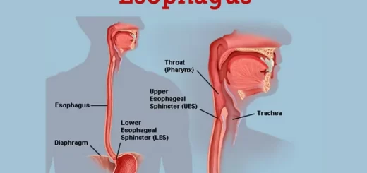
Histological organization of pharynx, Structure & function of the esophagus
Pharynx function, anatomy, location, muscles, structure & Esophagus parts
Mouth Cavity divisions, anatomy, function, muscles, Contents of Soft palate and Hard palate
Norma lateralis features & anatomy, Mandible surfaces, nerves & ligaments