Stomach parts, function, curvatures, orifices, peritoneal connections and Venous drainage of Stomach
The stomach digests food and sends it to your small intestine, It has three functions: The stomach temporarily stores food, It contracts and relaxes to mix and break down food, It produces enzymes and other specialized cells to digest food.
The stomach
The stomach is situated in the left hypochondrium, epigastric, and umbilical regions of the abdomen. It is a J-shaped organ and has two orifices, the cardiac and pyloric orifices, two curvatures, the greater and lesser curvatures, and two surfaces, an anterior and a posterior surface.
Parts of the stomach
- Fundus: is dome-shaped and projects upward and to the left of the cardiac orifice, It is usually full of gas.
- Body: extends from the fundus to a line extending from the incisura angularis on the lesser curvature to the prepyloric bulge on the greater curvature.
- pyloric part: it is the terminal part of the stomach. It has three parts (pyloric antrum, pyloric canal, and pyloric sphincter).
Curvatures of the stomach
- Lesser curvature: forms the right border of the stomach and extends from the cardiac orifice to the pylorus, The lesser omentum extends from the lesser curvature to the liver, It is related to the right and left gastric vessels.
- Greater curvature: forms the left border of the stomach and extends from the cardiac orifice to the pylorus. It gives attachment to gastrophrenic, gastrosplenic, and greater omentum from above downwards. It is related to the right and left gastroepiploic vessels.

Stomach parts
Orifices of the stomach
- Cardiac orifice: is 1 inch to the left of the median plane at the level of T.10, It is a physiological sphincter as it depends on the presence of mucosal folds, positive intra-abdominal pressure, and few fibers from the right crus of the diaphragm which encircle it.
- The pyloric orifice is 1/2 inch to the right of the median plane at the level of L.1 (transpyloric plane). It is a true anatomical sphincter as it contains circular muscle fibers.
Peritoneal connections of the stomach
- Lesser omentum.
- Greater omentum.
- Gastrosplenic ligament.
- Gatrophrenic ligament.
Lesser omentum
It is a fold of the peritoneum connecting the liver with the stomach.
Attachments
- To the porta hepatis and fissure for ligamentum venosum of the liver.
- To the lesser curvature of the stomach and 1st inch of the duodenum.
Contents:
- Right gastric vessels.
- Left gastric vessels.
- Portal vein.
- Hepatic artery.
- Bile duct.
- Lymph nodes.
- Sympathetic nerves.
- Extraperitoneal fat.
Greater omentum
It is a fold of the peritoneum connecting the greater curvature of the stomach with the anterior border of the pancreas. The transverse colon and mesocolon are attached to their posterior layers.
Attachments
- To the greater curvature of the stomach and first two inches of the duodenum.
- To the anterior border of panceas.
Contents:
- Right gastroepiploic vessels.
- Left gastroepiploic vessels.
- Lymph nodes.
- Sympathetic nerves.
- Extraperitoneal fat.
Gastrosplenic ligament
It is a short fold of the peritoneum connecting the spleen at its hilum with the posterior surface of the stomach close to the upper part of the greater curvature.
Contents:
- Short gastric vessels.
- Lymph nodes.
- Extraperitoneal fat.
Gastrophrenic ligament
It is a short fold of the peritoneum connecting the upper part of the back of the stomach with the diaphragm.
Relations of the stomach:
Relation of Anterior surface
- Hepatic area (liver).
- Anterior abdominal wall.
- Diaphragmatic area (Diaphragm).
Relation of Posterior surface (stomach bed)
It is related posteriorly to the lesser sac, left crus of the diaphragm, left suprarenal gland, the upper part of the left kidney, splenic artery, pancreas, transverse mesocolon transverse colon, and spleen separated from it by the greater sac.
Arterial supply to the stomach
These are derived from the branches of the coeliac trunk.
- The left gastric artery arises from the coeliac artery.
- The right gastric artery arises from the hepatic artery.
- Short gastric arteries arise from the splenic artery.
- The left gastroepiploic artery arises from the splenic artery.
- The right gastroepiploic artery arises from the gastroduodenal branch of the hepatic artery.
Venous drainage of the stomach
- The left and right gastric veins drain directly into the portal vein.
- The short gastric veins and the left gastroepiploic vein join the splenic vein.
- The right gastroepiploic vein joins the superior mesenteric vein, The right gastric vein is connected to the right gastroepiploic vein through the prepyloric vein of Mayo, This is an important landmark for the gastroduodenal junction.
Lymph drainage of stomach
The gastric lymph vessels drain ultimately into the celiac lymph nodes.
Nerve supply of the stomach
- Sympathetic: from celiac plexus around the celiac trunk. It causes relaxation of the wall and contraction of the pyloric sphincter. It carries also a pain sensation.
- Parasympathetic: from anterior and posterior gastric nerves (vagus), It is secretory to the glands of the stomach, inhibitory to the pyloric sphincter, and motor to the wall.
You can download Science online application on Google Play from this link: Science online Apps on Google Play
Regulation of stomach emptying, Causes of vomiting & Nervous pathway of reflex vomiting
Physiology & functions of Stomach, Composition of gastric secretion
Histological structure of stomach, Fundic glands of Stomach & Gastric musculosa
Constituents of the gastric juice, Gastric motility & types of movements occur in the stomach
Abdomen muscles, Blood Supply of Anterior Abdominal Wall & Rectus Sheath content
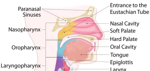
Histological organization of pharynx, Structure & function of the esophagus
Mouth Cavity divisions, anatomy, function, muscles, Contents of Soft palate & Hard palate