Temporal and infratemporal fossae contents, Muscles of mastication and Otic ganglion
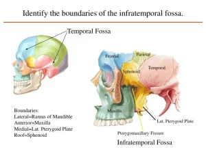
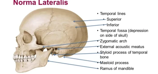
Temporal and infratemporal fossae are the interconnected spaces on the lateral side of the head, Their boundaries are formed by bone and soft tissues, The temporal fossa is superior to the infratemporal fossa, above the zygomatic arch, and communicates with the infratemporal fossa below through the gap between the zygomatic arch and the more medial surface of the skull.
Boundaries of Temporal and infratemporal fossae
The infratemporal fossa is a wedge-shaped space deep to the masseter muscle and the underlying ramus of the mandible laterally and the lateral wall of the pharynx medially.
Contents
2 Muscles: Medial and lateral pterygoids, Nerves: Mandibular, maxillary and Chorda tympani, 2 Vessels: Maxillary artery and Pterygoid plexus of veins and 1 ganglion: Otic ganglion.
Of the four muscles of mastication (masseter, temporalis, medial pterygoid, and lateral pterygoid) that move the lower jaw at the temporomandibular joint, one (masseter) is lateral to the infratemporal fossa, two (medial and lateral pterygoid) are in the infratemporal fossa, and one fills the temporal fossa (temporalis).
Muscles of mastication
There are four muscles of mastication:
- Temporalis
- Masseter
- Medial pterygoid
- Lateral pterygoid
Temporalis muscle
- Origin: Floor of temporal fossa and deep surface of temporal fascia, inferior temporal line.
- Insertion: Coronoid process of the mandible.
- Nerve supply: Deep temporal nerves (from the anterior division of the mandibular nerve).
- Action: Elevation (anterior fibers) and retraction (posterior fibers) of the mandible.
Masseter muscle
- Origin: Lower border and the inner surface of the zygomatic arch.
- Insertion: Lateral aspect of the ramus of the mandible Nerve
- supply: anterior division of the mandibular nerve.
- Action: Elevation and protraction of the mandible.
Lateral pterygoid muscle
- Origin:
- Upper head: from the infratemporal surface of the greater wing of the sphenoid bone.
- Lower head: from the lateral surface of the lateral pterygoid plate.
- Insertion: anterior aspect of the neck of the mandible and the articular disc of the temporomandibular joint.
- Nerve supply: anterior division of the mandibular nerve.
- Action: pulls the head of the mandible forward during the opening of the mouth (helps in the depression of the mandible), protracts the mandible, and side to side movement (chewing).
Medial pterygoid muscle
- Origin:
- Superficial head: from the maxillary tuberosity.
- Deep head: from the medial surface of the lateral pterygoid plate.
- Insertion: medial surface of the ramus of the mandible above the angle and below the mylohyoid groove.
- Nerve supply: The main trunk of the mandibular nerve.
- Action: helps in elevation of the mandible, protraction of the mandible, and side-to-side movement.
Mandibular nerve
Origin: it is the largest division of the trigeminal nerve. It has a small motor root and a large sensory root.
Course:
It enters the Infra-temporal fossa through the foramen ovale where both roots join each other to form the trunk of the nerve which is very short and rapidly divides below the foramen into anterior and posterior divisions.
I- Branches of the trunk:
- The nerve to the medial pterygoid muscle which supplies tensor palati and tensor tympani muscles passes through the otic ganglion without a relay.
- Nervous spinosus: Meningeal branch to meninges of middle cranial fossa.
Il- Branches of Anterior division: (All its branches are motor EXCEPT the buccal Nerve).
- Muscular branches: to masseter, lateral pterygoid, and temporalis.
- Buccal nerve: It supplies the skin covering the cheek. It pierces the buccinator to supply the mucous membrane of the mouth on its inner surface.
III- Branches of Posterior division: (All branches are sensory EXCEPT the mylohyoid nerve)
1. Inferior alveolar nerve:
It innervates the lower teeth, The inferior alveolar nerve and its accompanying vessels enter the mandibular foramen on the medial surface of the ramus of the mandible and travel anteriorly through the bone in the mandibular canal, Before it enters the mandibular canal it gives motor nerve to the mylohyoid muscle (which pierces the sphenomandibular ligament) and anterior belly of the digastric muscle, Adjacent to the first premolar tooth, the inferior alveolar nerve divides into incisive and mental branches:
- The incisive branch innervates the related teeth and gum.
- The mental nerve exits the mandible through the mental foramen and innervates the chin and lower lip.
2. Lingual nerve:
It originates in the infratemporal fossa and passes anteriorly into the floor of the oral cavity and continues forward on the medial surface of the mandible adjacent to the last molar tooth at the posterior end of the mylohyoid line (dangerous position). The lingual nerve then continues anteromedially across the floor of the oral cavity and ascends into the tongue on the lateral surface of the hyoglossus muscle. It supplies the mucous membrane of the floor of the mouth and carries general and taste sensations from the anterior two-thirds of the tongue.
3. Auriculo-temporal nerve: It arises by two roots that encircle the middle meningeal artery. It supplies the scalp over the temporal region and carries the parasympathetic supply to the parotid gland.
Otic ganglion
It is a parasympathetic ganglion in the infratemporal fossa below the foramen ovale medial to the main trunk of the mandibular nerve.
Roots:
- Sensory root: from the auriculotemporal branch of the mandibular nerve.
- Sympathetic root: from the plexus around the middle meningeal artery.
- Parasympathetic root: the fibres of this root arise from the inferior salivary nucleus and pass through the glossopharyngeal nerve. They leave the nerve in its tympanic branch; which reach the tympanic cavity where it breaks into the tympanic plexus. The fibers leave the plexus through lesser superficial petrosal nerve which reaches the infratemporal fossa through the foramen oval and then relay in the otic ganglion. (Only the parasympathetic root relays in the ganglion).
Branches:
- Parasympathetic postganglionic branches to the parotid gland.
- Sensory branches to the parotid gland.
- Sympathetic branches to blood vessels of the parotid gland. The ganglion is also traversed by the nerve to the medial pterygoid which passes through the ganglion without a relay to supply tensor palati and tensor tympani.
Maxillary artery
Origin: The maxillary artery originates from the external carotid artery within the substance of the parotid gland.
Course: The course of the artery is divided into 3 parts according to its relation with the lateral pterygoid muscle.
- First part: Medial to the neck of the mandible.
- Second part: On the lateral surface of the lateral pterygoid muscle.
- Third part: Passes between the two heads of the lateral pterygoid to reach the pterygopalatine fossa.
Branches:
1- Branches of 1st part: It gives the middle meningeal and inferior alveolar arteries and a number of smaller branches (deep auricular, anterior tympanic, and accessory meningeal).
2- Branches of 2nd part: It gives origin to deep temporal, masseteric, buccal, and pterygoid branches for medial and lateral pterygoid muscles, which course with branches of the mandibular nerve.
3- Branches of the 3rd part: (in the pterygopalatine fossa): It gives posterior superior alveolar, infra-orbital, greater palatine, pharyngeal, and sphenopalatine arteries, and the artery of the pterygoid canal. The infraorbital artery gives:
- Orbital branches to the orbit.
- Anterior superior alveolar artery.
- Terminal branches in the face (palpebral, nasal, and labial branches). Collectively, these branches supply much of the nasal cavity, the roof of the oral cavity, upper teeth, sinuses, oropharynx, and floor of the orbit.
Pterygoid Plexus of Veins
Site: It is a network of veins lying around and within the lateral pterygoid muscle.
Tributaries:
- Veins corresponding to the branches of the maxillary artery.
- Inferior ophthalmic vein through the inferior orbital fissure.
- Deep facial vein.
Drainage: The pterygoid plexus drains into the maxillary vein which unites with the superficial temporal vein to form the retromandibular vein.
Function: It acts as a peripheral heart to aid venous return by the pumping action of the lateral pterygoid.
Connections:
It communicates with:
- Anterior facial vein through the deep facial vein.
- Cavernous sinus via emissary veins passing through foramen ovale, foramen spinosum, and emissary sphenoid foramen.
- Inferior ophthalmic vein via a communicating vein passing through the inferior orbital fissure.
You can download Science online application on Google Play from this link: Science online Apps on Google Play
Histological organization of pharynx, Structure & function of the esophagus
Pharynx function, anatomy, location, muscles, structure & Esophagus parts
Mouth Cavity divisions, anatomy, function, muscles, Contents of Soft palate and Hard palate
Norma lateralis features & anatomy, Mandible surfaces, nerves & ligaments


