Skin appendages types, function (hair, nails, sweat glands and sebaceous glands)
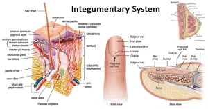
Skin appendages are epidermal and dermal-derived components of the skin that include hair, nails, sweat glands, and sebaceous glands. The skin, along with hair and nails, is the protective covering of the body, the skin prevents germs from entering the body and damaging the internal organs, the skin manufactures vitamin D when exposed to the sun’s ultraviolet rays, and vitamin D is an essential vitamin for healthy skin.
Skin appendages
Skin appendages are skin-associated structures, they serve a particular function including sensation, contractility, lubrication, and heat loss. Skin appendages (or adnexa) are derived from the skin, and they are adjacent to it, The skin supports the life of all other body parts and plays a role in maintaining the immune system.
Types of appendages include hair, glands, and nails. hairs are used for sensation, heat loss, filter for breathing & protection, arrector pilli (smooth muscles that pull hairs straight), sebaceous glands secrete sebum onto hair follicle, which oils the hair, sweat glands can secrete sweat with a strong odour (apocrine) or with a faint odour (merocrine or eccrine), and nails (protection).
Skin appendages function
Skin appendages are structures embedded within or projecting from the skin that perform various important functions. They play an important role in keeping our skin healthy and functioning properly. These include:
- Hair: Hair helps to insulate the body, protects the scalp from sunlight, and provides sensory input. It also traps sweat and dirt, helping to keep the skin clean.
- Nails: Nails protect the tips of the fingers and toes, and aid in grasping and scratching.
- Sweat glands: Sweat glands help regulate body temperature by producing sweat, which evaporates from the skin’s surface and cools the body. There are two main types of sweat glands: eccrine glands, which are found all over the body, and apocrine glands, which are found in areas like the armpits and groin.
- Sebaceous glands: Sebaceous glands produce sebum, an oily substance that lubricates the skin and hair, and helps to prevent them from drying out. Sebum also has some antimicrobial properties.
In addition to these main functions, skin appendages also play a role in:
- Sensation: Hair follicles and nerve endings in the skin help us to feel touch, pressure, and temperature.
- Protection: Hair, nails, and the outer layer of the skin (epidermis) help to protect the body from harmful substances, such as bacteria and viruses.
- Vitamin D production: The skin can produce vitamin D when exposed to sunlight.
Hair
Hairs are horny threads varying in length, thickness, color, and distribution according to the race, sex, and site of skin.
Histological features:
- The hair shaft develops from the hair follicle which is a cylindrical down growth of the epidermis into the dermis, hair follicle ends by the hair bulb.
- The hair bulb is invaginated from below by avascular connective tissue dermal papilla, which if destroyed, the hair follicle dies and no hair will grow again.
Nail
Nails are horny plates present on the dorsal surfaces of the terminal phalanges of fingers and toes, they consist of closely-packed hard keratin scales.
Sebaceous glands
These are simple branched alveolar, holocrine glands that develop from the upper third of the hair follicle; therefore, they are usually present where the hair is present. They are most numerous over the head region and the anogenital area.
Histological features:
1- The secretory portion:
- It is a pale flask-shaped structure formed of one or more alveoli with a common duct.
- The alveolus is lined with a layer of flattened germinal cells lying on a basement membrane, these cells proliferate by mitosis, enlarge by accumulating fatty material, become polyhedral, and are pushed to the center, where they degenerate and discharge sebum (holocrine mode), thus; sebum is a mixture of fatty substances, cell debris, and keratin.
2- The excretory duct is short and wide, it opens obliquely into the upper third of the hair follicle or directly into the epidermal surface. The duct is lined by stratified squamous epithelium.
The function of the sebaceous gland
Sebaceous glands secrete sebum which lubricates the skin surface and hair, also has a bactericidal effect. Sebaceous glands are relatively inactive until puberty when they are stimulated by the rising levels of sex hormones.
A disturbance in the normal flow of sebum is one of the reasons for the development of acne during adolescence, Acne is a chronic inflammation of obstructed sebaceous glands.
Sweat glands
They are simple coiled tubular glands, they are found deep in the dermis or the hypodermis, there are two types of sweat glands; the eccrine and the apocrine glands.
The eccrine sweat glands
They are distributed all over the body, they secrete a watery secretion rich in sodium chloride.
Histological features:
1- The secretory portion appears in histological sections in the form of small rounded acini with a narrow lumen and lined by stratified cuboidal epithelium composed of three cell types:
- The dark pyramidal cells secreting glycoprotein mucoid substance.
- The clear cuboidal cells secreting a watery secretion.
- The myoepithelial cells to move the secretion into the ducts.
2- The long excretory duct is lined by stratified cuboidal cells, It ascends in a helical course to the epidermis where it opens on the skin surface.
The apocrine sweat glands
They are present only in the skin of the axilla, areola of the breast, and perianal region, they begin their secretory function at puberty, their secretion is viscous and has a characteristic odor.
Histological features:
- The acini of the secretory portion are larger and of wider lumen than the eccrine glands, the secretory portion is composed of only two cell types; large cuboidal cells with apical secretory granules and myoepithelial cells.
- The excretory duct is lined by stratified cuboidal cells and opens into the hair follicle.
- The secretory cells undergo merocrine, not apocrine, secretion, thus; the glands are misnamed.
Functions of the integumentary system
- Protection of the body against mechanical, chemical, thermal, and bacterial agents, this occurs through the heavily keratinized cells of the stratum corneum.
- Protection of the body against water loss or gain (waterproof-barrier function), this occurs through the lipid-rich extracellular material in the stratum granulosum and corneum.
- Screening against sun ultraviolet radiation through melanin pigments.
- Perception of different stimuli from the environment through the presence of various receptors (sensory nerve endings) in the skin.
- Regulation of the body temperature:
– In hot weather, cooling of the body is enhanced by increased sweat secretion and evaporation with vasodilation of the dermal capillaries and closure of the arteriovenous (A-V) shunts, these mechanisms allow maximum cutaneous blood flow and heat loss.
– In cold weather, constriction of dermal blood vessels and opening of the A-V shunts to reduce cutaneous blood flow and reduce heat loss. - Excretion of nitrogenous products and sodium chloride in sweat.
- Formation of vitamin D in the epidermis (mainly in stratum basale and spinosum) when the skin is exposed to sunlight.
Integumentary system, Skin importance, layers, types and function
Embryonic connective tissue, Connective tissue proper, and Specialized connective tissue
Connective tissue cell types, function & structure, Resident cells, and Transient cells
Connective tissue structure, types, function, fibers, and ground substances
Tissue types, Epithelial tissue features, Covering and Glandular Epithelium
Carbohydrates importance, Types of Isomerism, Monosaccharides and Disaccharides


