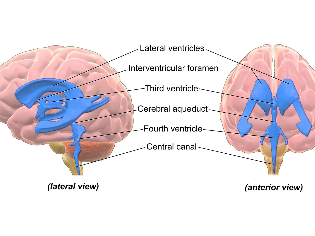
Ventricular system of the brain, Boundaries of 4 ventricles and pathway of Cerebrospinal (CSF)
The ventricular system of the brain consists of the intercommunicating 4 ventricles: The 2 lateral ventricles are C-shaped cavities; one for each cerebral hemisphere. The third ventricle is a slit-like cavity of the diencephalon. The fourth ventricle is the tent-like cavity of the hindbrain.
The lateral ventricle
It consists of the central part (body) and 3 horns; anterior, posterior, and inferior. Each lateral ventricle communicates medially with the 3rd ventricle through the interventricular foramen of Monro which lies at the junction between the body and the anterior horn of the lateral ventricle.
Anterior horn
It lies within the frontal lobe. It is limited anteriorly by the genu of the corpus callosum. It is continuous with the central part posteriorly at the interventricular foramen. It is related to:
- Parts of corpus callosum anteriorly, inferiorly, and superiorly.
- Head of caudate nucleus laterally.
Central part: it extends from the interventricular foramen till the splenium of the corpus callosum. It is related to:
- Body of corpus callosum superiorly.
- Body of caudate nucleus and thalamus inferiorly.
Posterior horn: it lies within the occipital lobe. It is related to the Tapetum of the corpus callosum and optic radiation laterally.
Inferior horn: it lies within the temporal lobe. It is the largest of the 3 horns. It is related to:
- The tail of the caudate and amygdaloid nucleus superiorly.
- Tapetum of the corpus callosum and optic radiation laterally.
Ventricular system of the brain
Third ventricle
It is the cavity of the diencephalon. It communicates with the lateral ventricle on each side through the interventricular foramen of Monro. The cavity of the third ventricle is traversed by the interthalamic adhesions.
Communications of the third ventricle
It communicates with:
- Lateral ventricles through the interventricular foramen of Monro.
- The fourth ventricle through the cerebral aqueduct of Sylvius.
The third ventricle has a roof, floor, 2 lateral walls, anterior wall, and posterior wall. The floor is formed by (from anterior to posterior):
- Optic chiasma.
- Tuber cinereum & infundibulum of pituitary gland.
- Mammillary bodies.
- Posterior perforated substance.
- Tegmentum of the midbrain.
Lateral wall is related to:
- Thalamus.
- Hypothalamus.
- Hypothalamic sulcus.
Fourth ventricle
It is a tent-like cavity of the hindbrain. It lies between the pons & medulla anteriorly and the cerebellum posteriorly
Boundaries of the 4 ventricles
Roof:
- Upper part: the superior medullary velum stretching in between the superior cerebellar peduncles.
- Lower part: Inferior medullary velum stretching in between the two inferior cerebellar peduncles.
Lateral wall:
- Upper part: superior cerebellar peduncles.
- Lower part: inferior cerebellar peduncles and gracile & cuneate tubercles.
Floor: It is rhomboidal in shape called homboid fossa. It is formed by the dorsal surface of the pons and medulla. Thus the floor of the fourth ventricle is divided into pontine and medullary parts separated by the stria medullaris. It communicates with:
- Subarachnoid space (through foramina of Luschka and Magendie).
- Third ventricle (through the aqueduct of the midbrain).
- The central canal of the spinal cord.
The cerebro-spinal (CSF)
CSF is a clear transparent fluid that fills all the cavities inside the brain ventricles and surrounding subarachnoid space.
Communications inside the ventricular system:
- The 2 lateral ventricles communicate with the third and ventricle through the interventricular foramina of Monro (one between each lateral ventricle and the third ventricle).
- The 3rd ventricle communicates with the 4th ventricle through the cerebral aqueduct (aqueduct of Sylvius).
- The 4th ventricle communicates with: The central canal of the spinal cord (downwards), and the subarachnoid space through; the 2 foramina of Luschka laterally and the foramen of Magendie at the midline.
The subarachnoid space:
It is space lies between the arachnoid and the pia maters.
Formation of the CSF: formed by the choroid plexuses of the lateral, 3rd and 4 ventricles.
Absorption: within the superior sagittal sinus through the arachnoid villi and granulations (herniation of arachnoid matter into dural venous-sinuses especially the superior sagittal sinus ”SSS”).
Pathway of the CSF
- From the lateral ventricles to the 3rd ventricle through the 2 interventricular foramina of Monro.
- From the 3rd to 4th ventricle through the cerebral aqueduct.
- From 4th ventricle to:
- Subarachnoid space through the foramina of Luschka and Magendie (behind the brain, then below and lastly around the supero-lateral surface).
- The central canal of the spinal cord. When the CSF reaches the top of the brain, it is absorbed from the subarachnoid space to the SSS through the subarachnoid villi and granulations. Part of CSF passes downward in the subarachnoid space around the spinal cord and is reabsorbed into the venous plexus surrounding the dura in the spinal cord.
You can follow Science Online on YouTube from this link: Science online
Cerebellar cortex structure, function, location, layers & fibers
Cerebellum function, structure, anatomy, location & blood supply
White matter of the brain function, structure and blood supply of the internal capsule
Anatomy of basal nuclei (basal ganglia) & Disorders of basal ganglia motor circuits
Function and Physiology of Thalamus, Hypothalamus & Limbic system
Diencephalon function, Thalamus, Metathalamus, Hypothalamus, Epithalamus & Subthalamus
Metabolism of the brain, Cerebrospinal fluid function, location & symptoms