Lymphatics of head and neck, superficial vessels, deep vessels and Abnormalities of branchial arches
The lymphatic system drains tissue fluid, plasma proteins, and other cellular debris back into the bloodstream, it is also involved in immune defense, The lymphatic vessels of the head and neck can be divided into two major groups; which are superficial vessels & deep vessels, Superficial vessels drain lymph from the scalp, face, and neck into the superficial ring of lymph nodes, The deep lymphatic vessels arise from the deep cervical lymph nodes, They converge to form the left and right jugular lymphatic trunks.
Lymphatics of head and neck
It is formed of 2 main systems:
Superficial group
Horisontal group:
- Submental.
- Submandibular.
- Pre-auricular (parotid).
- Post-auricular (mastoid)
- Occipital.
Vertical group:
- The midline group called anterior cervical along the anterior jugular vein.
- Two lateral groups are called superficial or lateral cervical lymph nodes: along the external jugular vein.

Lymphatics of head and neck
Deep group
Horizontal group:
Called (Waldeyer’s ring) and formed of:
- Lingual tonsils.
- Palatine tonsils.
- Tubal tonsils
- Pharyngeal tonsils.
Vertical group:
a. Along the midline includes:
- Infrahyoid
- Prelaryngeal
- Pretracheal
- Paratracheal
- Retropharyngeal
b. Two lateral groups along the internal jugular veins, includes:
- Upper deep cervical lymph nodes (one of them called jugulo-digastric).
- Lower deep cervical lymph nodes (one of them is called jugulo-omohyoid).
Branchial (pharyngeal) apparatus
The branchial arches are six arches, which develop in a successive manner from above down-ward around the pharynx (e. the first arch will appear first, followed by the second followed by the third and so on).
Each arch receives a separate branch from the aorta to supply it, The artery supplying the fifth arch degenerates early, so the 5th arch itself degenerates too, Now there are 5 arches named according to their number: 1st, 2nd, 3rd, 4th and 6th arch.
The first arch is also called the mandibular arch, the second is termed the hyoid arch. Each arch is horseshoe-shaped and formed of an outer layer of ectoderm, inner layer of endoderm separated by an intermediate layer of mesoderm, Each arch has an artery, nerve cartilage, and muscular component:
- On the ectodermal (outer) surface, each arch is separated from the next arch by a depression which is called a groove or cleft.
- On the endodermal (inner) surface, each arch is separated from the next one by a depression called a pouch.
Derivatives of branchial arches
The first or mandibular arch
It divides into small maxillary and large mandibular parts. Its nerve supply is the mandibular nerve. It contains cartilage called Meckle’s cartilage.
Derivatives of the first arch
- Skeletal derivatives: The maxillary part gives part of the maxilla, zygomatic bone, and temporal bone. The mandibular part gives two ossicles of the middle ear (malleus, and incus, anterior ligament of malleus, spheno-mandibular ligament and the mandible.
- Muscular derivatives: It gives the 4 muscles of mastication, tensor palati, tensor tympani, mylohyoid, and anterior belly of digastric muscle (all these muscles are supplied by the mandibular nerve).
The second arch (hyoid arch)
It is supplied by the facial nerve. Its cartilage is called Reichert’s cartilage.
Derivatives of the second arch
- Skeletal derivatives give the stapes (ear ossicle), styloid process of the temporal bone, stylohyoid ligament, lesser horn, and upper part of the body of the hyoid bone.
- Muscular derivatives: it gives the muscles of the face and scalp, platysma, posterior belly of the digastric, stylohyoid muscle, and stapedius muscle.
The third arch
It is supplied by the glossopharyngeal nerve.
Derivatives of the third arch
- Skeletal derivatives: it gives the greater horn & lower part of the body of the hyoid bone.
- Muscular derivatives: it gives only one muscle, which is the stylopharyngeus.
The fourth arch
This arch is supplied by the external laryngeal nerve (branch of the superior laryngeal of the vagus nerve).
Derivatives of the fourth arch
- Skeletal derivatives: thyroid cartilage.
- Muscular derivatives: cricothyroid muscle.
The sixth arch
It is supplied by the recurrent laryngeal nerve (branch from vagus nerve).
Derivatives of the sixth arch
- Skeletal derivatives: cricoid, arytenoid, cuneiform, and corniculate cartilages.
- Muscular derivatives: it gives all the intrinsic muscles of the larynx except the cricothyroid muscle.
Endodermal derivatives of the pharyngeal pouches
- The first pouch: it will form a diverticulum with a long stalk. The diverticulum will form the middle ear cavity, while the stalk will form the auditory tube.
- The second pouch: the endoderm after proliferation gives the palatine tonsil.
- The third pouch divides into dorsal and ventral wings. The dorsal wing will give the inferior parathyroid gland, while the ventral wing will give the thymus gland.
- The fourth pouch divides also into dorsal and ventral wings, The dorsal wing gives the superior parathyroid gland, while the ventral wing gives the ultimobranchial bodies.
Ectodermal derivatives of pharyngeal clefts
Four clefts are present between the branchial arches. They are lined by ectoderm, The first cleft penetrates the underlying mesoderm to form the external auditory meatus. The mesoderm of the second arch proliferates to form the operculum, This operculum grows caudally to overlap the third and fourth clefts. The operculum forms a space between it and the clefts. This space is called the cervical sinus. This sinus will disappear to give a smooth contour of the neck.
Abnormalities of branchial arches
- External cervical sinus: it is due to the failure of obliteration of the cervical sinus.
- Internal cervical sinus: it is a blind-ended pouch that opens into the pharynx.
- Branchial cyst: it is due to the closure of both ends of the sinus and a part of the middle remains.
- Branchial fistula: in this case, there is communication between the pharynx and the outside.
You can subscribe to science online on Youtube from this link: Science Online
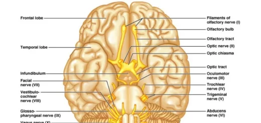
Cranial nerves types, Facial, Vestibulocochlear, Glossopharyngeal, Vagus, Accessory & Hypoglossal
Cranial nerves anatomy, function, Olfactory, Optic, Oculomotor, Trochlear, Trigeminal & Abducent
Triangles of the neck contents, Structure of Anterior & Posterior triangle of the neck
Facial muscles function, anatomy, arteries, veins, names & expressions
Skull function, anatomy, structure, views & Criteria of neonatal skull
Cranial cavity anatomy, function, structure, Dural folds, Cavernous sinus & Nerve supply of scalp