Nerves of forearm and hand, Nerve injuries types and causes
The forearm consists of the radius bone laterally and the ulna bone medially. The forearm serves as a bridge between the motion of the arm and the wrist and hand. The forearm nerves are responsible for innervating the muscles of the forearm. In addition to motor function. the nerves of the forearm offer afferent cutaneous sensation to the forearm, wrist, and hand.
Nerves of forearm and hand
The forearm nerves are complex due to the various nerve branches and the muscles that reside in the upper extremities. The nerves carry a lot of information such as electrical, biochemical (immune substances, growth factors, and hormones), and they participate in the metabolism of the tissue in which they pass.
Median nerve
Course:
- It enters the forearm by passing between the 2 heads of pronator teres.
- The median nerve then descends in the forearm between flexor digitorum superficialis and profundus.
- Two inches above the wrist, the nerve becomes superficial in position (critical position of the nerve).
- The nerve reaches the hand by passing deep to the flexor retinaculum.
- It then divides into medial and lateral terminal branches.
Branches:
- Muscular branches. For all superficial flexors of the forearm except flexors carpi ulnaris.
- Articular branches. To elbow and superior radioulnar joint.
- Anterior interosseus nerve. Supply all deep flexors of the forearm except the medial half of flexor digitorum profundus.
- Palmar cutaneous nerve. It passes superficial to flexors retinaculum to supply the lateral 2/3 of the skin of the palm.
- The lateral and medial terminal branches: giving rise to:
- Muscular branches to muscles of the thenar eminence and the lateral 2 lumbricals.
- Cutaneous branches to the skin of palmar aspect of the lateral 3 ½ fingers, nail beds, and dorsum of terminal phalanges of these fingers.
Ulnar nerve
Course:
- It enters the forearm by passing between the 2 heads of flexor carpi ulnaris.
- The ulnar nerve descends in the forearm between flexor carpi ulnaris and flexor digitorum profundus.
- In the lower part of the forearm, it becomes superficial covered only by skin and fasciae.
- In the lower part of the forearm, the nerve is closely accompanied by the ulnar artery.
- It enters the hand by passing superficial to the flexor retinaculum.
- It divides into superficial and deep branches.
Branches:
- Muscular branches. to flexor carpi ulnaris and medial ½ of flexor digitorum profundus.
- Articular branches to the elbow joint.
- Palmar cutaneous branch: to supply the skin of medial 1/3 of palm.
- Dorsal cutaneous branch: to the skin of medial 1/3 of the dorsum of the hand & medial 1 ½ fingers.
- Superficial terminal branch: to the palmaris brevis muscle and skin of medial 1 ½ fingers.
- Deep terminal branch: Supplies the following muscles:
- Hypothenar muscles(3).
- The third & fourth (medial 2) lumbrical muscles.
- All palmar interossei (4).
- All dorsal interossei (4).
- Adductor pollicis muscle.
Radial nerve
The radial nerve terminates in front of the lateral epicondyle by dividing into superficial and deep terminal branches.
The superficial branch
It continues under the cover of brachioradialis. Then it enters the dorsum of the hand. This superficial branch has no branches in the forearm. It supplies the skin of the lateral 2/3 of the dorsum of the hand and the posterior surface of the lateral 3 ½ fingers. The skin of the distal and middle phalanges of these fingers is supplied by the median nerve.
The deep branch (posterior interosseus nerve)
It pierces the supinator muscle to continue at the back of the forearm. The nerve supplies all the muscles at the back of the forearm (extensor group) except brachioradialis, extensor carpi radialis longus, and anconeus which are supplied by the radial nerve itself.
Nerve injuries
Brachial plexus – Upper roots (C5,6) injury
Causes:
- Increased angle between the head and the shoulder.
- Difficult delivery.
Effects: (Erb’sDuchenne palsy)
Motor effect:
- The Shoulder – adducted and medially rotated (arm hangs by the side).
- The elbow – extended.
- The forearm – pronated.
Sensory effect: Loss of sensation on the lateral side of the upper limb.
Brachial plexus – Lower Roots (C8, T1) injury
Causes:
- Excessive abduction of the arm.
- Cervical rib.
Motor effect:
- Opposite of writing position: Hyperextension of metacarpophalangeal joints + Flexion of interphalangeal joints (Complete claw hand = Klumpke’s palsy).
- Loss of abduction and adduction of fingers.
Sensory effects: Loss of sensation on the medial side of the upper limb.
Median nerve injury
Causes:
- Above the elbow: supracondylar fracture.
- Above the wrist: stab wound.
- Carpal tunnel syndrome: arthritis or synovitis
1- Injury above the wrist
Motor Effect:
- Ape’s hand deformity.
- Week thenar muscles.
- Loss of opposition of the thumb.
Sensory Effect: Lateral 2/3 of the palm and lateral 3.5 fingers (parathesia or anaesthesia).
2- Carpal tunnel syndrome
Motor Effect: Week thenar muscles. In neglected cases: as injury above the wrist.
Sensory Effect: Pins and needles sensation along with the distribution of the median nerve to the lateral three and half fingers. In neglected cases, there is a loss of sensation from the same fingers only.
3- Injury above the elbow
Motor Effect:
- Loss of pronation.
- Adduction of the wrist.
- Loss of flexion of interphalangeal joints of 2,3 digits (pointing index).
- + injury above the wrist.
Sensory Effect: As injury above the wrist.
Ulnar nerve injury
Causes:
- Above the elbow: Facture medial epicondyle.
- Above the wrist: Cut wounds.
Injury above the wrist
Motor effect:
- Partial claw hand.
- Loss of adduction and abduction of the fingers.
- Loss of adduction of the thumb.
- Flattening of the hypothenar eminence.
Sensory effect: Loss of sensation over medial 1/3 of the palm and medial 1.5 fingers.
Injury above the elbow
Motor effects:
- As above + less apparent clawing (ulnar paradox).
- Abduction of the hand.
Sensory effects: Loss of sensation over medial 1/3 of the hand and medial 1 ½ fingers.
Radial nerve injury
Causes
- Radial Nerve: In the axilla, pressure from the crutch or back of a chair. In the spiral groove, fracture of shaft of the humerus.
- Posterior Interosseous Nerve: Fracture of the head, neck, or the upper shaft of the radius. Downward dislocation of the head of the radius.
- Superficial Radial Nerve: Cut wounds.
Radial Nerve
Motor effects:
- Inability to extend the elbow.
- Inability to extend the wrist (Wrist Drop).
- Inability to extend the fingers (Fingers Drop).
Sensory effects:
Paresthesia over: Lower lateral surface of the arm. The posterior surface of the arm and forearm. Lateral 2/3 of the dorsum of the hand and posterior surface of the proximal phalanges of the lateral 3 ½ fingers.
Posterioi Interosseus Nerve
Motor effects:
- Inability to extend the fingers (Fingers drop).
- No Wrist Drop (intact extensor carpi radialis longus)
No sensory effects.
Superficial Radial Nerve
No motor effects.
Sensory effects: Paresthesia over lateral 2/3 of the dorsum of the hand and posterior surface of the proximal phalanges of the lateral 3 ½ fingers.
Forearm bones, anatomy, function & Skeleton of the hand
Flexors of forearm, Forearm muscles, structure, function & anatomy
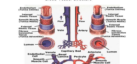
Blood vessels of forearm & hand, Veins and Lymphatics of the upper limb
Types of joints of upper limb, Ligaments & movements of the shoulder joint