Anatomy of brainstem, Features of medulla oblongata, pons and midbrain
The brainstem is the posterior stalk-like part of the brain, it connects the cerebrum with the spinal cord, In the human brain, It is composed of the midbrain, the pons, and the medulla oblongata, It plays an important role in the regulation of cardiac and respiratory function, consciousness, and the sleep cycle.
Anatomy of brainstem
The brainstem (or brain stem) is an important part of the brain, as the nerve connections from the motor and sensory systems of the cortex pass through it to communicate with the peripheral nervous system.
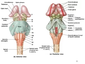
External features of the medulla oblongata
A. The ventral surface presents the following features
- Anteromedian fissure is an upward extension of the anteromedian fissure of the spinal cord, In the lower part of the medulla, the anteromedian fissure is traversed by the decussating fibers of the pyramidal tract (motor decussation).
- The elevation on each side of the anteromedian fissure is called a pyramid, The pyramid is formed by the corticospinal fibers before its decussation.
- The oval elevation lateral to the pyramid is called olive and is formed by the bulging of the olivary nuclei mainly the inferior olivary nucleus.
- The groove between the pyramid and the olive is called the anterolateral sulcus and it gives an exit for the rootlets of the hypoglossal nerve.
- The groove lateral to the olive is called the posterolateral sulcus. It gives exit to the rootlets of the glossopharyngeal, vagus, and cranial accessory nerves arranged from above downwards.
B. The dorsal surface presents the following features
The posteromedian sulcus is an upward continuation of the posteromedian sulcus of the spinal cord, The longitudinal elevation lateral to the posteromedian sulcus is called gracile fasciculus as it overlies the gracile tract, The upper end of the gracile fasciculus expands to form the gracile tubercle which overlies the gracile nucleus.
The longitudinal elevation lateral to the gracile fasciculus is called cuneate fasciculus as it overlies the cuneate tract, The upper end of the cuneate fasciculus expands to form the cuneate tubercle which overlies the cuneate nucleus, The cuneate tubercle is present lateral to and extends to a higher level than the gracile tubercle.
The ridge lateral to the cuneate tubercle is the inferior cerebellar peduncle, The area between the two inferior cerebellar peduncles forms the lower part of the floor of the fourth ventricle, This area is triangular in shape and is bounded above by the stria medullaris, This area presents an inverted V-shaped depression called inferior fovea which divides it into three trigones from medial to lateral:
- Hypoglossal trigone that overlies the hypoglossal nucleus
- The vagal trigone overlies the dorsal nucleus of the vagus.
- Vestibular trigone that overlies the inferior vestibular nucleus and part of the medial vestibular nuclei.
External features of the pons
A. The ventral surface presents the following features:
- It is convex from side to side; marked by prominent transverse ridges.
- Laterally, it is continuous with the middle cerebellar peduncles on each side. It presents a median groove called a basilar groove as it lodges the basilar artery.
- The trigeminal nerve emerges from the middle part of the pons at its junction with the middle cerebellar peduncle, The abducent nerve emerges at the lower border of the pons, between it and the pyramid, The facial and vestibulo-cochlear nerves also emerge at the lower border of the pons; between it and the olive (The facial nerve is medial to the vestibulo-cochlear).
B. The dorsal surface presents the following features:
- It forms the upper part of the floor of the fourth ventricle, This part presents a depression called superior fovea that separates the eminentia medialis (medial eminence) from the upper vestibular area.
- The lower part of the medial eminence presents a prominent elevation called facial colliculus, This colliculus is produced by the abducent nucleus surrounded by the facial nerve fibers, The upper vestibular area is produced by the lateral and superior vestibular nuclei as well as part of the medial vestibular nucleus.
External features of the midbrain
The midbrain has a narrow lumen called cerebral aqueduct (aqueduct of Sylvius), A coronal plane passing through the cerebral aqueduct divides the midbrain into two divisions; cerebral peduncles and the tectum:
A. The cerebral peduncle is differentiated into 3 parts
- Crus cerebri (Basis pedunculi): this is formed of bundles of nerve fibers descending from the cerebral cortex to lower levels of the brainstem and spinal cord; these are corticospinal, corticobulbar, and corticopontine fibers.
- Substantia nigra: it is a lamina of pigmented grey matter containing melanin pigment, lying dorsal to the crus cerebri.
- Tegmentum: this is the posterior part of the cerebral peduncle and is continuous inferiorly with the tegmentum of the pons. The red nucleus is located in the tegmentum of the midbrain on either side of the midline.
B. Tectum
This is the smaller dorsal part of the midbrain, The tectum is formed of four knob-like elevations called colliculi or corpora quadrigemina, They are arranged as two superior and two inferior colliculi, Each colliculus gives rise to a brachium from its lateral side, The superior brachium connects the superior colliculus with the lateral geniculate body.
The inferior brachium connects the inferior colliculus with the medial geniculate body, The transverse sections of the brainstem at its different levels are correlated to the shape and size of grey and white matter in these levels.
Interpeduncular fossa
It is a rhomboidal space at the junction of the midbrain and the base of the cerebrum.
Boundaries:
- Anteriorly: optic chiasma.
- Anterolaterally: optic tracts.
- Posterolaterally: crura cerebri.
- Posteriorly: upper border of the pons.
Contents:
- Oculomotor nerves: each nerve emerges immediately medial to the corresponding crus.
- The posterior perforated substance: this is a layer of grey matter in the angle between the crura cerebri. It is pierced by the central branches of the posterior cerebral arteries.
- The mammillary bodies: these are pairs of small white spherical bodies that protrude from the ventral surface of the hypothalamus immediately anterior to the posterior perforated substance.
- The tuber cinereum: it is a slightly raised area of grey matter between the mammillary bodies and the optic chiasma. The infundibulum arises from the tuber cinereum; it is a narrow stalk that connects the hypothalamus to the pituitary gland.
Functional classification of cranial nerves
Cranial nerves can be classified into:
- Sensory: olfactory, optic, vestibulo-cochlear.
- Motor: oculomotor, trochlear, abducent, accessory, hypoglossal.
- Mixed: trigeminal, facial, glossopharyngeal, vagus.
Deep origin and functional types of cranial nerve nuclei are represented in:
The oculomotor nerve (III):
- Oculomotor nucleus (GSE).
- Edinger Westphal nucleus (GVE).
Trochlear nerve (IV): Trochlear nucleus (GSE).
Abducent nerve (VI): Abducent nucleus (GSE).
Trigeminal nerve (V):
- Motor nucleus of trigeminal (SVE).
- Main sensory nucleus of trigeminal (GSA, fast pain).
- Spinal nucleus of trigeminal (GSA, slow pain & temperature).
- Mesencephalic nucleus (GSA, proprioception).
Facial nerve (VII):
- Motor facial nucleus (SVE).
- Superior salivary nucleus (GVE).
- Solitary nucleus (GVA, SVA).
Glossopharyngeal nerve (IX):
- Nucleus ambiguus (SVE).
- Inferior salivary nucleus (GVE).
- Solitary nucleus (GVA, SVA).
- Spinal nucleus of trigeminal (GSA).
Vagus nerve (X):
- Nucleus ambiguus (SVE).
- Dorsal vagal nucleus (GVE).
- Solitary nucleus (GVA, SVA).
- Spinal nucleus of trigeminal (GSA).
The accessory nerve (XI):
- Cranial part: from nucleus ambiguus (SVE).
- Spinal part: from upper 5 or 6 spinal cervical segments (GSE).
The hypoglossal nerve (XII): Hypoglossal nucleus (GSE).
You can follow science online on YouTube from this link: Science online
You can download Science online application on Google Play from this link: Science online Apps on Google Play
Stretch reflex (myotatic reflex) types, properties, Muscle tone, muscle spindles and Clonus
Physiology of central human reflexes, Types & properties of Spinal cord reflexes
Physiology & function of the spinal cord, Lateral & medial brainstem pathway
Histological organization spinal cord, Relation between spinal & vertebral segments
Blood supply of CNS (central nervous system), Carotid system & Circle of Willis


