Nucleus components, function, diagram and classification of chromosomes
The nucleus is the largest membranous organelle of the cell. It contains the chromosomes together with the machinery for DNA replication and RNA transcription and processing. The nucleus directs the process of protein synthesis in the cytoplasm by informational macromolecules (RNAs) formed in the nucleus and transported to the cytoplasm.
The nucleus
Usually, each cell has a single nucleus. Sometimes, liver cells are binucleated. Skeletal muscle fibers (cells) are multinucleated, whereas mature RBCs have no nuclei.
Components of the interphase nucleus
- The nuclear envelope.
- The chromatin, genetic material.
- The nucleolus.
- The nucleoplasm (nuclear sap).
The nuclear envelope
It consists of two parallel membranes, outer and inner separated by the perinuclear cisterna. It is perforated by the nuclear pores that provide a channel through which the nucleus and cytoplasm communicate and exchange substances.
The outer membrane
It is continuous with the rough endoplasmic reticulum. It is covered with ribosomes adhering to its outer surface. These ribosomes synthesize the transmembrane proteins of the nuclear membranes.
The inner membrane
It is supported at its inner surface by the lamins forming the nuclear lamins.
The function of the nuclear lamina
- Supports the nuclear envelope.
- Influences chromosome distribution and function within the nucleus.
The nuclear pores
They are perforations in the nuclear envelope where the outer and inner nuclear membrane fuse. These pores are not uniformly distributed and vary in number from few to several thousands in metabolically-active cells. The nuclear pores provide a bidirectional channel through which the nucleus and cytoplasm communicate and exchange substances e.g. ribosomal subunits.
The chromatin
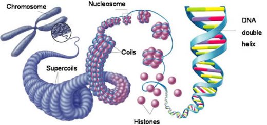
Chromatin is formed of deoxyribonucleic acid (DNA) conjugated with histone proteins. Because of its contents of nucleic acids, the nuclear chromatin strongly binds basic dyes such as hematoxylin. DNA is extensively-packaged in chromatin as:
- A segment of the DNA double helix is wrapped about two times around a central core of eight histone proteins to form the structural unit of chromatin, a nucleosome. Each nucleosome particle is separated from the next by a region of the linker DNA.
- Repeating nucleosomes with intervening “linker” DNA form a 10nm fiber, known as “beads on a string”.
- This chain of nucleosomes is usually packed together to form a 30nm fiber.
- Higher orders of packaging, eventually give the compact structure 700nm seen in the metaphase of the dividing cell known as the chromatid of a chromosome.
According to its appearance in the interphase nucleus, two forms of chromatin are known :
Heterochromatin (condensed chromatin), which represents the inactive part of chromatin. It may be able to transform into euchromatin when needed.
LM picture
It appears as dense basophilic clumps.
EM picture
It appears as condensed filaments or granules distributed in the following sites:
- Nucleolar-associated heterochromatin, around the nucleolus.
- Peripheral heterochromatin, at the inner nuclear membrane associated with the nuclear lamina.
- Heterochromatin islands, swimming in the nuclear sap.
Euchromatin (extended chromatin), which represents the active part of chromatin, i.e. this chromatin is stretched out so that the genetic information in the DNA can be transcribed.
LM picture
It appears as lightly-stained basophilic areas.
EM picture
It appears as dispersed filaments or granules.
The proportion between euchromatin and heterochromatin differs from one cell to another according to its functional activity. Active cells e.g. dividing cells and those with high synthetic activity have a lightly-stained euchromatic nucleus with a prominent nucleolus. During cell division, chromatin is condensed and organized into the chromosomes.
Metaphase chromosome
A typical metaphase chromosome is formed from two chromatids held together at the centromere. Each chromatid is formed of a single DNA molecule that contains many genes.
Classification of chromosomes
The human somatic cell contains 46 chromosomes organized as 23 pairs. Karyotyping is the systemic arrangement of the chromosomes during metaphase into groups of homologous pairs. According, chromosomes are organized into 22 homologous pairs of autosomes and one pair of sex chromosomes.
The sex chromosomes
- They are concerned with sex characteristics. They are identical female (XX) and non-identical in males (XY).
- In females with normal karyotype (44 autosomes + XX), one X chromosome of the sex pair is heterochromatic in the interphase nucleus.
- It is present in the form of a dense chromatin mass called “Barr body”. It can be identified in neutrophils, attached to one of the nuclear lobes in the form of a drumstick mass.
The nucleolus
It is a spherical body within the nucleus with no surrounding membrane. Metabolically-active cells have highly-basophilic large or multiple nucleoli. It is the site of formation (transcription) of ribosomal RNA. These rRNAs are packaged with their associated proteins to form the ribosomal subunits that are exported to the cytoplasm via the nuclear pores to start the process of protein synthesis.
The nucleoplasm (nuclear matrix)
It is a colloidal protein solution that provides a medium for the rapid diffusion of metabolites and for the movement of different kinds of RNAs towards the nuclear pores.
Cytoplasmic organelles, Ribosomes & Endoplasmic reticulum function, structure & definition
Cell Structure, the function of Golgi apparatus, Endosomes & Lysosomes
The function of Cytoplasmic organelles, Mitochondria, Peroxisomes & Cytoskeleton
Structure of Cytoplasm, The function of centrosome & Cytoplasmic inclusions
Histology, Molecular structure of the cell membrane, Cell function & structure
Regulation of the cell cycle, DNA synthesis phase, Interphase & Mitosis