Nervous system (Central nervous system, Peripheral nervous system and Autonomic nervous system)
Nervous system consists central nervous system (Brain and Spinal cord), the peripheral nervous system (Cranial nerves and Spinal nerves), Autonomic nervous system (Sympathetic nervous system and parasympathetic nervous system).
Central nervous system
As previously mentioned the central nervous system is divided into the brain and spinal cord.
Brain
It is the major part of the central nervous system and its weight is about: 350 grams at birth, and 1400 grams in an adult man, It occupies a bony space called the brain case or the skull (cranium), It is surrounded by three membranes called the meningies which are responsible for the protection and nutrition of brain cells, 12 pairs of cranial nerves are connected to the brain.
These membranes are :
- The dura mater which lines the skull.
- The pia mater which is in direct contact and adheres to the brain.
- The arachnoid which is in between the other two membranes and contains a transparent fluid to protect the brain from mechanical trauma.
The main components of the brain are Forebrain (Brain cortex, Thalamus, Hypothalamus), Midbrain, and Hindbrain (Cerebellum, Pons Varolii, Medulla oblongata).
The structure and function of each part of the brain:
Forebrain
It represents the largest part of brain , It consists of two cerebral hemispheres and the cerebral cortex, the Thalamus, and the Hypothalamus.
Two cerebral hemispheres and cerebral cortex
Two big lobes separated by a big fissure and attached to each other by a big bundle of nerve fibers, Each lobe is called the cerebral hemisphere, The cortex of each lobe (cerebral cortex) is characterized by the presence of depressions of different depths called fissures and grooves and in between there are folds.
Each cerebral hemisphere is divided into many lobes, which are the frontal lobe, parietal lobe, occipital lobe, and temporal lobe, The 5th lobe of cerebral cortex can not be seen by the naked eye as it is covered by the fronted and partial lobes.
Functions of cerebral cortex:
- The fronted lobe contains centers of voluntary movements (motor centers) and centres of memory and speech.
- Parietal lobe controls many sensory functions and contains centres of sensation of heat, cold, pressure and touch (somatic sensations from the skin).
- Occipital lobe contains centres of vision .
- The temporal lobe contains centres of smell and taste, and also centres of hearing.
Thalamus
It is an important centre for the coordination of different sensations (except the smell) that reach the cortex.
Hypothalamus
It controls the different reflex actions and contains centres of : (Hunger, Satiety, Thirst, Body temperature regulation, Sleep).
Midbrain
It is the smallest part of the brain, It represents a connection between the forebrain and hindbrain, It contains centers of Equilibrium (keeping the body balance), Hearing, and Vision, It regulates many reflexes as those related to hearing.
Hindbrain
It consists of Cerebellum, Pons Varolii, and medulla oblongata, Cerebellum is situated in the posterior region and consists of three lobes, It keeps the balance and equilibrium of the body in association with the inner ear and muscles.
Pons Varolii and medulla oblongata perform the following functions :
- Transmission of nerve impulses between the spinal cord and different brain regions.
- The medulla oblongata contains vital centres such as those of respiration, swallowing, vomiting, cough, sneezing, and blood vessel movement.
Spinal cord
It exists inside a canal in the vertebral column called the neutral canal , It extends from the medulla oblongata in the form of cylindrical cord , Its length is about 45 cm long , It is hollow from the inside due to containing a central canal .
There are two fissures (dorsal and ventral) extend along the midline which divide the spinal cord incompletely into two halves, It is covered by three membranes (meningies), which are Dura mater , Pia mater, and Arachnoid.
Structure of the spinal cord
It consists of two layers :
- The outer white layer (white matter) is formed from nerve fibers.
- The inner grey layer (grey matter) is H-shaped with two dorsal horns and two ventral horns formed from nerve cells with their dendrites and neuroglia.
Its function :
- White matter transmits impulses from the different body parts to the brain and vice versa.
- Grey matter is the main centre of reflex action, as it contains thousands of reflex arcs.
Peripheral nervous system
It connects the central nervous system with all parts of the body, It consists of a network of nerves distributed all over the body, which includes Cranial nerves and Spinal nerves.
Cranial nerves
Number: there are 12 pairs connected to the brain, Types: sensory (containing sensory fibers only), motor (containing motor fibers only), or mixed nerves.
Mixed nerves are nerves carry the impulses from the receptors to the brain and from the brain to the effector organs, so, they are sensory and motor nerves together.
Spinal nerves
Number: 31 pairs connected to the spinal cord and they are mixed nerves with both sensory and motor fibers, Spinal nerves are divided into :
- Cervical nerves: 8 pairs.
- Thoracic nerves: 12 pairs.
- Lumbar nerves: 5 pairs.
- Sacral nerves: 5 pairs.
- Coccigeal nerves: 1 pair.
Types: all spinal nerves are mixed (sensory and motor nerves), Each spinal nerve originates from the spinal cord by two roots (dorsal and ventral).
The dorsal root carries sensory nerve fibers, It transmits the impulses from the receptors to the spinal cord, then to the brain.
The ventral root carries motor nerve fibers, It transmits the impulses from the brain and spinal cord to the responding (effector) organs such as muscles and glands.
Reflex arc (Reflex action)
The Reflex arc is the unit of nervous activity, The majority of nervous functions can be analyzed into a group of reflex actions, The reflex action consists of at least two nerve cells:
- Sensory nerve cell (Afferent).
- Motor nerve cell (Efferent).
The majority of reflex actions of five elements:
- Receptor (Sense organs).
- Afferent (Sensory) neuron.
- Connector (Intermediate) neuron.
- Efferent (Motor) neuron.
- Effector (Responding) organ.
Types of reflex arc:
- Voluntary (Somatic) reflex arc: The effector organ is the skeletal muscle.
- Involuntary (Autonomic) reflex arc: The effector organ is an involuntary muscles or glands or heart.
Autonomic nervous system
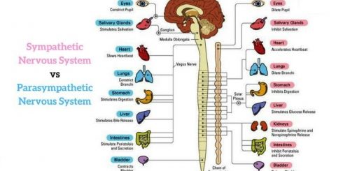
It regulates the different involuntary activities such as contraction of the cardiac muscles and smooth (involuntary) muscles, and secretion of the body glands, It consists of sympathetic nervous system and parasympathetic nervous system.
Sympathetic nervous system
The nerve fibers of this system arise from the thoracic and lumbar regions of the spinal cord, This system is important as an emergency system, as it enables the body to confront emergency situations.
Parasympathetic nervous system
The nerve fibers of this system arise from the brain stem and sacral region of the spinal cord.
Most of the internal parts of the body receive nerve fibers related to both the sympathetic and parasympathetic systems, and in most cases, the effect of one system antagonizes the effect of the other.
The effect of sympathetic and parasympathetic systems on some parts of the body
Heart: The sympathetic system increases the heartbeat rate and force of contraction, Parasympathetic system decreases the heartbeat rate and force of contraction.
Blood vessels: The effect of sympathetic NS is vasoconstriction ( contraction ) of blood vessels of the skin, viscera, salivary glands, brain, external genetalia, and lungs, Effects of Parasympathetic NS is vasodilation (Relaxation) of blood vessels of salivary glands and external genetalia.
Alimentary canal: The effect of sympathetic NS is relaxation of the wall of the stomach, intestine, and colon, The Effect of Parasympathetic NS is contraction of the wall of the stomach, intestine, and colon.
Respiratory system: The effect of sympathetic NS is dilation (Relaxation) of bronchioles and decreases their secretions, The Effect of Parasympathetic NS is constriction ( contraction ) of bronchioles and increases their secretions .
Urinary bladder: The effect of sympathetic NS is the relaxation of the wall of the urinary bladder, The Effect of Parasympathetic NS is the contraction of the wall of the urinary bladder.
Glands: Salivary glands (Sympathetic system stimulates little secretion of saliva, Parasympathetic system stimulates large secretion of saliva), Gastric juice of stomach (Sympathetic system inhibits the secretion, Parasympathetic system stimulates the secretion).
Liver: Sympathetic system breaks down the glycogen and increases the glucose level in the blood, Parasympathetic system decreases the glucose level in the blood.
Pancreas: The sympathetic system inhibits the secretion of enzymes, Parasympathetic system stimulates the secretion of enzymes.
Adrenal medulla: The sympathetic system stimulates the secretion of an adrenaline hormone (epinephrine) which increases blood pressure, heart rate, and the glucose level of blood, there is no parasympathetic fibers connection.
Autonomic nervous system, Reflex action types and Autonomic ganglia function
Nervous system in man, Nerve cells types and Nature of nerve impulse
Functions of Sympathetic nervous system and Role of the sympathetic in emergencies