Physiology and function of the spinal cord, Lateral and medial brainstem pathway
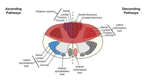
The functions of the spinal cord are transmitting the nerve impulses for movement, sensation, pressure, temperature, pain, and more, The spinal cord transmits nerve signals from the motor cortex to the body, and from the afferent fibers of the sensory neurons to the sensory cortex, It is a center for coordinating many reflexes and it contains reflex arcs that can control reflexes.
Physiology of the spinal cord
The spinal cord is the location of groups of spinal interneurons that make up the neural circuits known as central pattern generators, These circuits are responsible for controlling motor instructions for rhythmic movements such as walking.
Functions of the spinal cord
The spinal cord has the following functions:
- It transmits sensory signals from body parts to the cortex through the different ascending tracts.
- It transmits motor signals from the cortex to the muscles of the body and the face through the descending tracts.
- It performs reflexive actions.
- It contains preganglionic neurons of the autonomic nervous system.
Physiology of the motor descending tracts
The motor descending tracts can be functionally divided into two major groups:
- Pyramidal tracts: these tracts originate in the cerebral cortex, carrying motor fibers to the spinal cord and brainstem. They are responsible for the voluntary control of the muscles of the body and face. They comprise the corticospinal (ventral and lateral) and corticobulbar tracts.
- Extrapyramidal tracts: these tracts originate in the brainstem, carrying motor fibers to the spinal cord. They are responsible for automatic (subconscious) muscle control to support muscle tone, balance, posture, and equilibrium. They receive input from premotor areas and the cerebellum.
These tracts terminate in the ventral horn anterior horn cells (AHCs) of the spinal cord either directly or indirectly through interneurons. It should be noted that alpha motor neurons present in the ventral horn are topographically organized, with the cells controlling the distal portions of the limbs represented most laterally, and the cells controlling the proximal portions represented more medially. The ventral horn also contains the smaller gamma motor neurons.
I. Pyramidal tracts
1. The corticospinal tracts
About 60% of the corticospinal tract fibers have their cell bodies in the primary motor cortex, in the precentral gyrus of the frontal lobe, and in the premotor area. Primary and secondary somatosensory cortical areas located in the parietal lobe give rise to about 40% of the fibers of the corticospinal tract.
Axons of the corticospinal tract leave the cerebral cortex in the fan-shaped corona radiata and then they narrow as they descend to pass through the internal capsule, and then continue their descent through the ventral part of the midbrain, pons, and medulla.
In the medulla, the fibers collect forming the medullary pyramid. In the lower medulla, 80% of the fibers cross to the opposite side. (motor decussation) and continue in the contralateral side of the spinal cord forming the lateral (crossed) corticospinal tract terminating on the alpha motor neurons reaching distal muscles contralaterally and are involved in controlling skilled movements.
The remaining (20% of the fibers descend on the same side of the spinal cord forming the ventral (uncrossed) corticospinal tract, but about 14% of these fibers cross to the contralateral side only at the segmental level where they terminate on alpha motor neurons supplying proximal and axial muscles.
The remaining 6% of the fibers continue on the same side and supply proximal and axial muscles on the same side of the body. This means that the ventral corticospinal tract reaches proximal muscles bilaterally. Only a small percentage of the corticospinal tract fibers are directly. synapse on the alpha motor neurons, with the majority terminating on interneurons.
2. Corticobulbar tracts
The corticobulbar tracts originate from the face motor area and descend as collaterals from the corticospinal tract to supply muscles of the face, head, and neck. Corticobulbar fibers terminate on the brainstem motor nuclei bilaterally except the lower part of the facial nucleus and hypoglossal nucleus which receive fibers only from the opposite side.
The corticobulbar fibers which supply the extraocular muscles originate from the frontal eye field area and terminate on the motor cranial nerve nuclei III, IV, and VI cranial nerve and are termed corticonuclear tract.
Effect of lesion of pyramidal tract
Strokes of the motor cortex or corticospinal tracts are common. Their immediate consequences are paralysis of the fine movements on the contralateral side.
II. Extra-pyramidal tracts
The extrapyramidal tracts perform the following functions:
- They are responsible for the control of muscle tone, posture, and equilibrium.
- They are responsible for gross coordinated movements involving groups of muscles.
These tracts are divided, based on their location in the brainstem into:
- The lateral brainstem pathways: include the rubrospinal tract.
- The medial brainstem pathways: include the tectospinal, reticulospinal, and vestibulospinal tracts.
The lateral brainstem pathway
Rubrospinal tract
It originates from the red nucleus of the midbrain, The axons of these neurons cross the midline in the midbrain and descend to the contralateral spinal cord. They terminate mainly on interneurons in the cervical cord which in turn project to the alpha motor neurons for control of distal limb muscles (flexor muscles of the arm and forearm but not of individual digits).
Function: the function of this tract is to facilitate limb flexors.
The medial brainstem pathways
1. The tectospinal tract
It originates from the superior colliculus in the midbrain. The axons of this tract cross the midline and descend in the anterior part of the spinal cord to terminate at cervical segments.
Function: this tract is excitatory to neck extensors, to control of postural reflexes by directing head movements in response to visual and auditory stimuli.
2. Lateral reticulospinal tract
It originates from the inhibitory (medullary) reticular formation of the brainstem and terminates at all levels of the spinal cord mainly on the opposite side on the gamma motor neurons.
Function: it inhibits the tone of extensor muscles (axial and proximal limb muscles).
3. Medial reticulospinal tract
It originates from the facilitatory (pontine) reticular formation in the brainstem, descends on the same side, and terminates at all segments of the spinal cord mainly on the gamma motor neurons.
Function: it facilitates muscle tone of the antigravity and extensor muscles to maintain the body posture against gravity.
4. The lateral vestibulospinal tract
This tract arises from neurons of the lateral vestibular nucleus in the pons, the fibers in this tract are uncrossed and descend the entire length of the spinal cord.
Function: it activates the alpha motor neurons innervating extensor muscles of the trunk and ipsilateral limb to maintain the upright posture.
5. The medial vestibulospinal tract
This tract arises from the medial vestibular nuclei in the medulla and pons, descends in the cervical spinal cord, and terminates on the alpha motor neurons that innervate neck and upper limb muscles.
Function: the main function of this tract is to adjust the position of the head in response to vestibular stimuli.
Properties of the extrapyramidal tracts (compared to the pyramidal tract)
- Its pathway from the cortex to the spinal cord involves multiple neurons and synapses (not one neuron as in the pyramidal).
- They do not occupy the pyramid of the medulla oblongata.
- Only some of its tracts cross while many others descend directly (in contrast to the pyramidal which almost entirely crosses).
- It functions during the first year of life i.e, earlier than the pyramidal that functions later on in life.
Physiology of central human reflexes, Types & properties of Spinal cord reflexes
Histological organization spinal cord, Relation between spinal & vertebral segments
Features of synaptic transmission, Mechanism of sensitization & long term potentiation
Synaptic transmission steps, Synapses types & Nature of the postsynaptic change
Blood supply of CNS (central nervous system), Carotid system & Circle of Willis
Skull function, anatomy, structure, views & Criteria of neonatal skull
You can follow science online on YouTube from this link: Science online
You can download Science online application on Google Play from this link: Science online Apps on Google Play


