Functions of Lymphatic system, Structure of Lymph nodes, Spleen and Tonsils
The lymphatic system consists of the lymph nodes, spleen, thymus as well as the lymphatic tissue found in the small intestine (Peyer’s patches) and throat (adenoid tonsils, palatine & tubal tonsils), It is a part of your immune system, It has many functions, It can protect your body from illness-causing invaders, maintain body fluid levels, absorb digestive tract fats and remove cellular waste. Blockages, diseases or infections can affect your lymphatic system’s function.
Functions of the Lymphatic System
It consists of a complex network of lymphoid organs, lymph nodes, lymph ducts, lymph tissues, lymph capillaries, and a network of lymphatic vessels that carry lymph and other substances throughout the body. The lymphatic system can produce immune cells (such as lymphocytes, monocytes, and antibody-producing cells called plasma cells), It can absorb fatty acids and subsequent transport of fat, chyle, to the circulatory system.
The lymphatic system can remove excess fluids from body tissues, this process is crucial because water, proteins, and other substances are continuously leaking out of tiny blood capillaries into the surrounding body tissues, when the lymphatic system didn’t drain the excess fluid from the tissues, the lymph fluid would build up in the body’s tissues, and they would swell.
Lymph nodes
These are kidney-shaped bodies, which filter the lymph. They are located along the course of the lymphatic vessels. The lymph node is indented at the hilum where arteries enter while veins and efferent lymphatic vessels leave the lymph node. In the cut section, the lymph node consists of an outer dense cortex and an inner pale medulla.
Histological structure of the lymph node
Stroma
The lymph node is surrounded by a fibroelastic capsule that sends many perpendicular septa (trabeculae) that divide the cortex into several compartments. On reaching the medulla, these trabeculae run in different directions, anastomosing with each other. Reticular connective tissue network supporting the parenchymal cells and the lymph sinuses in its meshes.
Parenchyma
Cortex can be divided into two regions which are the outer cortex and inner cortex. The outer cortex is situated under the capsule. It consists of the cortical lymphoid nodules separated by the cortical lymph sinuses. The cortical nodules are rounded or oval dense lymphoid nodules formed by B lymphocytes and macrophages. When the lymph node is activated by the lymph-borne antigens, the B lymphocytes at the center of the nodule will develop into larger, less closely-packed lymphoblasts forming pale central areas called germinal centers.
The result of the germinal center formation is the production of an expanded population of B memory cells and plasma cells. The cortical lymphoid nodules are either primary (without germinal center) or secondary (with the germinal center). Inner cortex (thymus-dependent zone) or the paracortical area consists mainly of T lymphocytes which are not arranged as nodules.
The medulla: The dense lymphoid tissue in the medulla is in the form of wavy columns of cells, and the medullary cords. they branch and anastomose and are separated by medullary lymph sinuses. The medullary cords are formed of densely-packed B lymphocytes, plasma cells, and macrophages. They have no germinal centers. The loose lymphoid tissue is formed of loosely scattered lymphocytes, plasma cells, and macrophages with lymph sinuses surrounding the dense cortical lymphoid nodules and medullary cords.
Lymph circulation in the lymph node
Many afferent lymphatic vessels cross the capsule and pour lymph into the subcapsular sinuses. Lymph is drained to the cortical sinuses then into the medullary sinuses. The medullary sinuses converge towards the hilum where they drain the filtered lymph into the efferent lymphatic vessels.
The spleen
The spleen is the largest lymphoid organ in the body. Its function is to filter the blood. Histologically, the spleen is formed of stroma and parenchyma.
Histological structure of the spleen
Stroma
The spleen is surrounded by a thin capsule covered by peritoneum (mesothelial cells). At the hilum, the capsule is thickened and sends trabeculae that run in the splenic parenchyma in different directions carrying along with them the blood vessels. The capsule and the trabeculae consist of collagen, elastic, and numerous smooth muscle fibers. Reticular connective tissue network supporting the parenchymal cells and the blood sinusoids in its meshes. It is denser in the white pulp than in the red pulp.
Parenchyma: subdivided into white and red pulps
White pulp (The Malpighian corpuscles) is formed of lymphoid nodules, scattered in the substance of the spleen. These lymphoid nodules are traversed by the central artery or follicular artery which is always eccentric in position. The areas around the central arteries are called periarterial lymphoid sheaths (PALS) which are thymus-dependent areas consisting predominantly of T-lymphocytes. The lymphoid nodules are formed mainly of B lymphocytes & macrophages. They may contain germinal centers formed of lymphoblasts & plasma cells which then migrate to the red pulp.
The red pulp is formed of a network of branching and anastomosing blood sinuses (splenic blood sinusoids) separated from each other by the splenic cords or Billroth’s cords. The splenic cords are cellular masses of varying thicknesses and shapes, and consist of erythrocytes, macrophages, lymphocytes, plasma cells, and granulocytes.
The blood sinuses of the spleen are characterized by the presence of slits between the endothelial cells, so worn out or defective erythrocytes and platelets are severely damaged when they pass through these slits and phagocytosed by the macrophages associated with the sinusoidal walls.
The marginal zone
The marginal zone is the area around the margins of the lymphoid nodules between the red and white pulps. It is important because it is the site where:
- The circulating T & B lymphocytes enter into their specific location in the splenic parenchyma.
- Blood-borne antigens have their first access to the parenchyma of the spleen thus initiate the immune response and are attacked by the splenic macrophages.
The tonsils
Tonsils are organs composed of aggregations of incompletely- encapsulated lymphoid tissue. They form a ring surrounding the pharynx that lies beneath the epithelium at the beginning of the digestive and respiratory tracts. They include the palatine tonsils, the pharyngeal tonsil, and the lingual tonsils.
The palatine tonsils
The two palatine tonsils are located in the lateral walls of the oropharynx. Each tonsil is characterized by:
- The anterior and medial surfaces of the tonsil are covered by non-keratinized stratified squamous epithelium which dips inside the parenchyma to form primary and secondary crypts.
- The lumen of the crypts may contain desquamated epithelial cells, lymphocytes, and bacteria.
- Lymphoid nodules (primary and secondary) are present beneath the epithelium and around the crypts.
- Loose lymphoid tissue is present in between the lymphoid nodules. It consists of loosely-arranged lymphocytes, plasma cells, macrophages, and neutrophils.
- No lymph or blood sinuses are present.
- The lateral and posterior surfaces are covered by a connective tissue capsule that separates the tonsil from adjacent organs and acts as a barrier against the spreading of tonsillar infections in follicular tonsillitis. several mucous acini are present within the connective tissue capsule.
Lymphatic system structure, Function of Thymus, Vascular supply & blood-thymus barrier
Immune system structure, function, cells & Types of body defense mechanisms
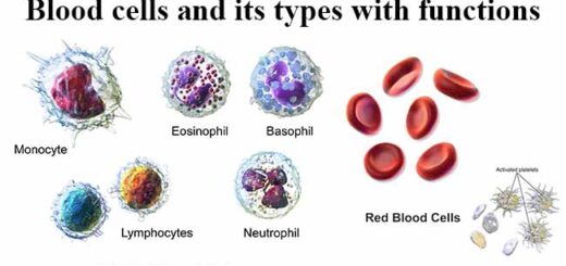
White Blood cells structure, function, types and How they are formed in the body
White Blood cells structure, function, types and How they are formed in the body
Red blood cells (Erythrocytes) structure & function, Myeloid tissue & Bone marrow
Carbohydrate Metabolism, Importance & Hormonal regulation of glycolysis