Nephroblastoma (Wilm’s tumor), Risk factors of Urothelial tumors (Tumors of transitional epithelium)
Wilms tumor is a malignant embryonal neoplasm derived from primitive nephrogenic blastemal cells which may differentiate into the epithelial and mesenchymal components, It is the most common renal neoplasm of childhood, It is the third most common solid (non-hematologic) cancer in children younger than 10 years of age. Age. 2-5 years, It occurs in sporadic and familial forms.
Nephroblastoma (Wilm’s tumor)
Morphology:
Grossly: The tumor appears as a large solitary well-circumscribed mass; however, 10% of cases are either bilateral or multifocal. Cut section is soft homogeneous and tan to grey with occasional foci of hemorrhage and necrosis.
Microscopically:
Tumors are characterized by attempts to recognizable attempts to recapitulate different stages of nephrogenesis. Classically the tumor is triphasic and composed of primitive blastema cells, Epithelial cells and stromal cells (although the percentage of each component is variable).
- Blastemal component, sheets of small blue round cells.
- Epithelial component. is in the form of abortive tubules or glomeruli.
- Stromal component, fibrous or myxoid.
Approximately 5% of tumors contain foci of anaplasia and carry a poor prognosis.
Clinical features
It manifests itself by:
- The large palpable abdominal mass which may extend across the midline and down into the pelvis (the main presentation).
- Less often: Fever, abdominal pain, Hematuria, intestinal obstruction as a result of pressure from the tumor.
Methods of spread
Local spread to the surrounding structures due to early invasion of the capsule, Bloodborne metastases to the lung, bone, and brain, Lymphatic spread to the regional lymph nodes.
Prognosis
The prognosis for Wilms tumor generally is very good, and excellent results are obtained with a combination of nephrectomy and chemotherapy, Diffuse anaplasia is associated with an adverse prognosis.
Urothelial tumors (Tumors of transitional epithelium)
The entire urinary collecting system from the renal pelvis to the urethra is lined by transitional cells (urothelium), Most urothelial tumors occur in the bladder, and Urothelial tumors above the bladder are relatively uncommon. Most urothelial tumors are malignant, but their aggressiveness and prognosis vary depending on microscopic grade and tumor stage. Most urothelial tumors are transitional cell carcinoma; however rare cases of squamous cell carcinoma and adenocarcinoma are also reported.
Tumors of the urinary bladder
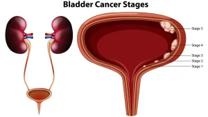
Most of them are transitional cell carcinoma (urothelial carcinomas), Squamous cell carcinoma occurs on top of squamous metaplasia (as in cases of bilharziasis), Rare cases of adenocarcinoma are also reported, Bladder carcinoma is most common in older males Age, most common between 50 and 80 years of age, Gender more common in men than in women, Painless hematuria is a common presenting symptom of the bladder cancer and requires clinical investigation by cystoscopy and/ or urine cytology analysis to rule out urothelial neoplasia.
Risk factors
Cigarette smoking (one of the most important risk factors).
- Industrial exposure to azo dyes.
- Various occupational carcinogens e.g. arylamines.
- Schistosoma hematobium infection predisposes to squamous cell carcinoma in endemic areas.
- Drugs such as analgesics and cyclophosphamide.
- Prior radiation therapy (cervical, prostatic or rectal cancer).
- Genetic alterations.
Transitional tumors
Classification:
According to WHO (2004), transitional tumors are classified according to invasion (tumor stage) into:
- Invasive urothelial carcinoma.
- Superficial Non-invasive tumors.
- Non-invasive flat urothelial tumors (CIS).
- Non-invasive papillary urothelial tumors.
The most important prognostic factor in non-invasive papillary urothelial neoplasms is their grade, which is based on both architectural and cytologic features. The grading subclassifies tumors as follows:
- Papilloma (rare).
- Papillary urothelial neoplasm of low malignant potential (rare) (PUNLMP).
- Low-grade papillary urothelial carcinoma.
- High-grade papillary urothelial carcinoma.
1. Non-invasive urothelial tumors
2. Invasive Urothelial Carcinoma
The extent of invasion and spread (TNM staging) at the time of initial diagnosis is the most important prognostic factor, Invades the lamina propria (non-muscle invasive) or extends more deeply into underlying muscle (muscle invasive), May be associated with precursor lesions (high-grade papillary urothelial cancer or CIS), Almost all invasive urothelial carcinomas are high-grade, The stage also determines treatment modality; with the invasion of the muscularis propria layer being an indication for radical cystectomy or radiation therapy with neoadjuvant or adjuvant chemotherapy.
Methods of Spread of Bladder Carcinoma
- Local to the surrounding structures.
- Lymphatic spread to lymph nodes.
- Blood-borne metastases.
Clinical features
- Painless hematuria.
- Dysuria.
- Hydronephrosis and/ or hydroureter (due to obstruction of vesicoureteric orifices).
You can subscribe to Science Online on Youtube from this link: Science Online
You can download Science Online application on Google Play from this link: Science Online Apps on Google Play
Neoplastic diseases of the urinary tract, Tumors of the kidney and Renal cell carcinomas
Urinary tract obstruction symptoms, causes and obstructive lesions of urinary tract
Urinary bladder structure, function, Control of micturition by Brain & Voluntary micturition
Urinary passages function, structure of Ureter, Urinary bladder & Uvulae vesicae
Urine formation, Factors affecting Glomerular filtration rate, Tubular reabsorption & secretion
Histological structure of kidneys, Uriniferous tubules & Types of nephrons
Functions of Kidneys, Role of Kidney in glucose homeostasis, Lipid & protein metabolism


