Sense of vision, Refractive media of the eye, Corneal reflex and Pupillary pathways
Sense of vision is one of the most important sensory functions, It serves as the basis for our perception of the external world, About 70% of all sensory receptors are in the eyes, and nearly half of the cerebral cortex is involved in some aspect of visual processing.
Refractive media of the eye
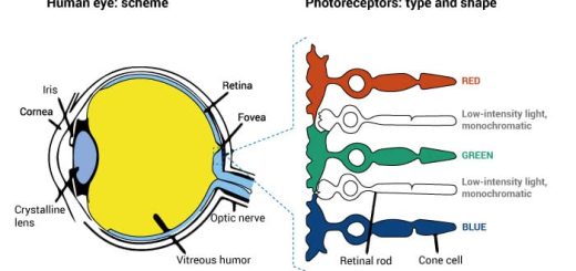
They are the structures that focus light rays entering the eye on the retina. The refractive power of 4 & 2 equal 7 diopters but they are usually negligible. The total refractive power of the human eye will be approximately 60 diopters when the lens is at rest i.e. looking for objects at a distance of 6 meters or more.
Since the refractive index of air is 1.0 and the differences between the indices of the cornea, aqueous and vitreous humor are little, the chief refractive media of the eye are the cornea and the lens.
The cornea is a refractive medium of the eye
It is the most important part of the refractive power of the eye; it provides about 42 diopters (about 2/3) of the total dioptric power of the eye, This is due to the refractive index of the cornea is markedly different from that of the air, The curvature of the cornea is great (it acts as a powerful convex lens).
The corneal reflex
The cornea plays an important role in eye protection. The corneal reflex protects the eye from the entrance of foreign bodies. This is a reflex closure of the eye on touching the cornea with any foreign body.
Pathway of the corneal reflex
- Afferent: sensory nerve fibers of the ophthalmic division of the trigeminal nerve pass to the trigeminal ganglia then to the trigeminal nucleus.
- Center: facial nucleus in the pons.
- Efferent: facial nerve.
- Effector: orbicularis oculi muscle which closes the eyelids.
The lens
The lens provides about 18 diopters (about 1/3) of the total refractive power of the optical system of the eye at rest (without accommodation). However, it can increase its power to enable the person to see near objects by accommodation.
The stimulus for accommodation:
- An object at infinity (Le. more than 6 meters from the eye) emits parallel rays which are refracted by the cornea and lens to form a clear focused image on the retina without any effort of accommodation.
- An object at a distance of 6 meters in front of the eye will have its focus still on the retina without the need for accommodation.
- The objects placed at a distance less than 6 meters in front of the eye without accommodation have focus behind the retina and its image will be blurred, In this case, if accommodation for near vision takes place, the lens increases its curvature to increase its refractive power and bring divergent light rays from near objects into a clear focus on the retina, This occurs by contraction of the ciliary muscle.
- In young individuals, the change in the shape of the lens may add about 14 diopters to the refractive power of the eye through accommodation.
- Accommodation requires increasing the optical power of the lens to focus near objects on the retina
Mechanism of accommodation
The main mechanism of accommodation is ciliary muscle contraction, In an unaccommodated state, resting tension on the zonule at the lens equator holds the lens in a relatively flattened state, When the ciliary muscle contracts, the resting zonular tension is released, and the lens is allowed to round up through the force exerted on the lens substance by the lens capsule, The lens anterior and posterior curvatures increase to increase the optical power of the lens.
The pupillary pathways
The function of the pupil is to control the amount of light that reaches the retina. It is controlled by the parasympathetic fibers (causing constriction) and sympathetic fibers (causing dilatation). The two most important pupillary reflexes are the near reflex and the light reflex.
The near reflex
The near reflex starts when the eyes are directed from a distant to a near object. The reflex is Initiated by the blurring of the retinal image. The near reflex has three components:
- Accommodation: contraction of the ciliary muscle leads to an increase of the refractive power of the lens, bringing the image to the retina.
- Convergence: contraction of the medial recti of both eyes, bringing the images of the near object on the foveae.
- Miosis: constriction of the pupils increases the depth of focus that is, the distance within which objects are seen without blurring.
Pathway of the near reflex
The stimulus: is the blurred image.
The receptors: are the photoreceptors (rods and cones).
The afferent: impulses travel from the retina through the optic nerve, optic chiasma, optic tract, lateral geniculate body, and optic radiation to the visual cortex. The visual cortex is connected to the frontal cortex. From there, the cortical fibers descend via the internal capsule to the (center) oculomotor nuclei in the midbrain. Some of these descending fibers synapse with parasympathetic nuclei (Edinger-Westphal nuclei) of the oculomotor nerve on both sides.
The efferent: the parasympathetic preganglionic fibers travel through the oculomotor nerves to the ciliary ganglia, where they synapse. The postganglionic fibers pass through the short ciliary nerves to the effector (ciliary muscle and constrictor pupillae muscle) to produce the response (accommodation and miosis respectively). Some of the descending cortical fibers go to the somatic part of the oculomotor nucleus then to the oculomotor nerve to contract both medial recti (causing convergence).
Accommodation is mediated by the parasympathetic fibers of the oculomotor nerve, So, the parasympathomimetic drugs (e.g. pilocarpine) induce accommodation, and parasympatholytic medications (e.g. atropine block accommodation, The drugs that relax the ciliary muscle are called cycloplegics.
The effect of age on accommodation: the nearest point to the eye at which an object can be brought into clear focus by accommodation is called the near point of vision. With advancing age, there is a decline in accommodation power due to hardness in the lens substance.
So, the near point of vision recedes throughout life from 9 cm at the age of 10 to about 83 cm at the age of 60. This condition is called presbyopia. As a result of this recession, close work becomes difficult at the age of 45-40 unless vision is corrected by reading glasses that use convex lenses.
The light reflex
When light falls on one eye its pupil constricts (the direct light reflex). At the same time, the pupil of the other unstimulated eye also constricts (the indirect or consensual light reflex). Under normal conditions, constriction of both pupils is of the same magnitude and it is proportional to the intensity of illumination. If light falls on both eyes simultaneously, the constriction of the pupils is greater than if the same intensity of light falls on one eye.
Pathway of the light reflex
The stimulus: The light reflex is initiated by stimulation of the visual receptors by light.
The afferent: the impulses are relayed to the bipolar cells to the ganglion cells of the retina. The axons of ganglion cells form the optic nerve. Partial crossing of the optic nerve fibers occurs at the optic chiasma, to form the optic tract (i.e. fibers coming from the nasal part of the retina cross in the chiasma to reach the optic tract of the opposite side, temporal fibers go to the optic tract of the same side).
The center: the light reflex fibers leave the optic tract before it reach the lateral geniculate body to relay in the pretectal nuclei of the midbrain. From each pretectal nucleus, impulses pass through the tectonuclear tract to Edinger-Westphal nuclei of the oculomotor nerve on both sides (the tectonuclear tract passes near the aqueduct of Sylvius).
The efferent: preganglionic fibers pass through both oculomotor nerves to relay in the ciliary ganglia.
The effectors: postganglionic fibers enter the eye through the short ciliary nerves, to end in the sphincter pupillae muscles of both eyes causing constriction of both pupils (the response is miosis in both eyes).
The consensual light reflex is explained by the partial decussation at the optic chiasma and by bilateral innervation of the Edinger-Westphal nuclei from each pretectal nucleus. The pathway of light reflex is subcortical and can be retained when there is a bilateral cortical lesion causing blindness.
Emergency room physicians routinely assess the pupillary light reflex because it is useful for assessing brainstem functions. Lack of pupillary light reflex or an abnormal reflex can be caused by optic nerve damage, oculomotor nerve damage, brainstem death or depressant drugs as barbiturates.
The lesion in the pretectal area near the aqueduct Sylvius due to syphilis, diabetes, or brain tumor produces Argyll Robertson pupil where the light reflex is lost due to destruction of tectonuclear tracts, but the near response is present because its pathway is intact.
You can subscribe to Science Online on YouTube from this link: Science Online
You can download the Science Online application on Google Play from this link: Science Online Apps on Google Play
Retina function, anatomy, layers, and accessory structures of the eye
Eyes structure, Histological organization of the fibrous and vascular coast of the eye
Visual system, Bony orbit anatomy, contents & Nerves, Muscles & movements of Eyeball
Retinal receptors, Photochemistry of vision, Steps of phototransduction in rods & Retinal output
Nystagmus types, causes, symptoms Development of CNS & Abnormalities of Spinal cord