Cytoplasmic organelles, Ribosomes and Endoplasmic reticulum function, structure and definition
Cytoplasmic organelles are metabolically-active structures carrying out specific essential functions. Most of the organelles are membranous organelles that are bounded by /or formed membranes that are similar to the cell membrane, these are the nucleus, endoplasmic reticulum, Golgi apparatus, transport vesicles, endosomes, lysosomes, mitochondria, and peroxisomes. The non-membranous organelles are also present, these are ribosomes, centrosome, and the cytoskeleton.
Ribosomes
Ribosomes are granules of nucleoproteins, ribosomal RNA (rRNA) conjugated with proteins. They are present in all cells especially abundant in cells where protein synthesis is taking place. Each ribosome is formed of two subunits, a small subunit, and a large subunit. The rRNAs of both subunits are synthesized inside the cell nucleus within its nucleolus.
Ribosomal-associated proteins are synthesized in the cytoplasm and then enter the nucleus and associate with rRNA. Ribosomal subunits then leave the nucleus, via the nuclear pores, to enter the cytoplasm. The small and large subunits are present in the cytosol individually and do not form a ribosome until protein synthesis begins.
LM picture
Ribosomes, when present in large amounts they cause cytoplasmic basophilia due to their contents of the nucleic acids.
EM picture
Ribosomes are small electron-dense granules, their appearance depends on their types and functions:
- Free ribosomes: they may be Solitary particles scattered in the cytoplasm that do not synthesize protein but act as a reserve. Aggregated (polysomes): in clusters of 10 or more connected by a single strand of mRNA. Polysomes are responsible for the synthesis of cytosolic protein, required for use within the cell and destined to remain in the cytosol, such as in dividing cells and growing cells e.g. synthesis of hemoglobin in developing red blood cells and contractile protein in muscle cells.
- Attached ribosomes: these are polysomes that become attached to the outer membrane of the endoplasmic reticulum making its surface rough.
Endoplasmic reticulum (ER)
The endoplasmic reticulum forms the most extensive membrane system in the cytoplasm. The ER has two types:
- Rough endoplasmic reticulum (rER).
- Smooth endoplasmic reticulum (sER).
Both types from a single membrane system but their histological appearance, relative proportions, and functions vary in different cell types.
Rough endoplasmic reticulum (rER)
LM picture
When present in large amounts they cause cytoplasmic basophilia due to their attached ribosomes.
EM picture
It consists of interconnected parallel flattened sacs called cisternae. It appears rough because its cytosolic surface is studded with ribosomes, resting by their subunit on the membranes of ER. It is continuous with the outer membrane of the nuclear envelope. The lumen contains flocculent material that represents a newly-formed protein.
The function of Rough endoplasmic reticulum
- Synthesis of secretory proteins e.g. hormones and enzymes.
- Synthesis of lysosomal proteins, enzymes, and proteins inserted into the cytoplasmic membranes.
- Post-translational modification of the newly-formed protein including folding, sulfation, and initial glycosylation.
- Transport the newly-synthesized protein to the Golgi body by transport vesicles.
Abundant in liver cells and protein-synthesizing and secreting cells e.g. pancreatic acini, fibroblasts, and plasma cells.
Smooth endoplasmic reticulum (sER)
LM picture
- It consists of a close network of interconnected branching tubules and vesicles.
- The membranes have a smooth surface as it lacks the attached ribosomes.
- The membranes of the sER are continuous with that of rER.
The function of Smooth endoplasmic reticulum
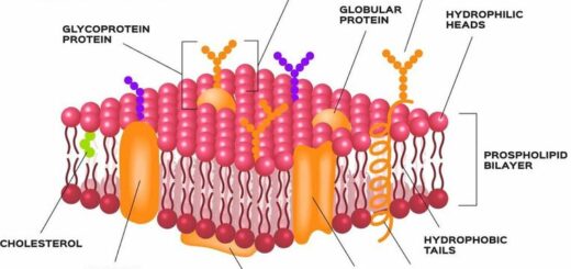
- Synthesis of membrane lipids, phospholipids, and cholesterol.
- Synthesis of stored hormones.
- Synthesis of glycogen in the liver.
- Detoxification and conjugation of toxic substances e.g. alcohol and drugs.
- Regulation of calcium ions during muscle contraction. sER in muscles is called sarcoplasmic reticulum.
Abundant in liver cells and steroid-secreting cells e.g. the adrenal cortex, testis, and ovary.
Cell Structure, the function of Golgi apparatus, Endosomes & Lysosomes
Function of Cytoplasmic organelles, Mitochondria, Peroxisomes & Cytoskeleton
Nucleus components, function, diagram & classification of chromosomes
Histology, Molecular structure of the cell membrane, Cell function & structure
Types of Transport through cell membranes, Active transport, Simple & Facilitated diffusion
Body fluid exchange, The electrical characteristic of the cells, Osmosis & Filtration
Vesicular transport of Macromolecules across the cell membrane, Endocytosis & Exocytosis