Cell Structure, the function of Golgi apparatus, Endosomes and Lysosomes
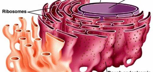
Cytoplasmic organelles are suspended in the cytoplasm of the cell, Each type of organelle has a definite structure & a specific role in the function of the cell. Cytoplasmic Organelles are the membrane-bound structures in a cell, such as a nucleus, Other organelles are located in the cytoplasm such as mitochondria, chloroplasts, endoplasmic reticulum, Golgi apparatus, peroxisomes, lysosomes, vacuoles & glyoxisomes.
Golgi apparatus
Golgi apparatus is found in all cells and is well-developed in the glandular secretory cells.
LM picture
It is not stained with H & E in routine histological sections. However, in H & E-stained sections of the cells synthesizing and secreting protein, the Golgi apparatus is well-developed and its site could be seen as a negative Golgi image, a clear pale area near the nucleus against the basophilic cytoplasm e.g. in plasma cells. It can be seen in histological sections with silver stain.
EM picture
It consists of a collection of flattened, curved, membrane-bounded saccules or cisternae called the Golgi stacks. Each stack consists of 4-6 cisternae. The cisternae are narrow throughout most of their length but slightly expanded at the ends. The Golgi stack is cup-shaped with a convex surface (cis face) and a concave surface (trans face):
- The cis face is convex in shape and lies near to the rER. It is also called a forming face or immature face because it is the site where small transport vesicles containing the newly-formed proteins from the rER enter the Golgi apparatus for further processing.
- The trans face is concave in shape and is also called a secretory face or mature face because it is the site where the modified protein is packaged and released from the Golgi in large secretory vesicles.
- The medial compartment is formed of the central cisternae of the stack between the cis and the trans compartments.
The function of the Golgi apparatus
Post-translational modifications of proteins previously-synthesized in rER through removal, addition or modification of sugars. Packaging of different proteins in the membrane-bounded vesicles. Sorting and targeting of vesicles to the right destination:
- Formation of lysosomes (in clathrin-coated vesicles) to remain inside the cell.
- Formation of secretory granules for exocytosis.
- Membrane recycling.
Endosomes
Endosomes are a system of vesicles and tubules involved in the endocytotic pathway. They are divided into two compartments:
Early endosomes
- They are formed at the periphery of the cell as a part in the pathway of the receptor-mediated endocytosis.
- They contain the receptors-ligands complex.
- The membrane of the endosomes actively pumps H+ ions into its interior. This lowers the pH of endosomes to less than 6.
- This acidity helps in the uncoupling of the receptors and the ligands.
- The receptors recycle to the cell membrane and the ligands move to the late endosomes.
Late endosomes
- They are irregular vesicles located deep within the cytoplasm near the Golgi complex. They receive the ligands from early endosomes and have acidic pH 5.5.
- They also receive clathrin-coated vesicles containing lysosomal enzymes from the Golgi complex. These enzymes become active at the acidic pH of the late endosome.
- The lysosomal enzymes in the late endosome begin to degrade the ligands accompanied by a further decrease in the internal pH which then “mature” to form lysosomes.
Lysosomes
They are membrane-bounded organelle containing about 40 types of acid hydrolytic digestive enzymes, including proteases, nucleases, phosphatases, and lipases. They are responsible for the intracellular digestion of different materials. Lysosomes are thought to be produced by a gradual maturation process as they are assembled by fusion of the clathrin-coated vesicles coming from the Golgi complex with late endosomes.
Lysosome has a surrounding membrane with unique phospholipids that prevents the leak out of the enzymes to the cytoplasm and also protects the membrane from hydrolysis by its own enzymes. If a lysosome leaks its contents, the released enzymes would be inactive because of the neutral pH of the cytoplasm.
LM picture
They can be recognized by several histochemical methods used to demonstrate the lysosomal enzymes.
EM picture
Lysosomes vary in their number in different cell types, they are abundant in phagocytic cells. Lysosomes are heterogeneous in shape and the appearance of their interior. Some are electron-dense, others show electron-lucent areas.
Pathways for intracellular digestion by lysosomes
Different pathways deliver material for digestion within the lysosomes, depending on the nature of the digested material.
- Extracellular small particles are internalized by pinocytosis and receptor-mediated endocytosis. They follow the endocytotic pathway through early endosomes then late endosome where the endocytosed materials are degraded by the lysosomal hydrolases.
- Extracellular large particles such as bacteria, cell debris, and other foreign materials are engulfed in the process of phagocytosis. A phagosome is formed when the material is internalized within the cytoplasm, subsequently fuses with a late endosome containing lysosomal enzymes.
- Intracellular particles such as worn-out or malfunctioning organelles are removed by a process called autophagy. The process begins with the enclosure of this organelle by membranes from sER, creating an autophagosome, which then fuses with a late endosome.
- The hydrolytic enzymes digest most of the content of the lysosomes. Any indigestible substances remain in lysosomes forming residual bodies.
- In long-lived cells, accumulated residual bodies indicate cellular aging and are called lipofuscin pigments.
Cytoplasmic organelles, Ribosomes & Endoplasmic reticulum function, structure & definition
Function of Cytoplasmic organelles, Mitochondria, Peroxisomes & Cytoskeleton
Nucleus components, function, diagram & classification of chromosomes
Histology, Molecular structure of the cell membrane, Cell function & structure
Types of Transport through cell membranes, Active transport, Simple & Facilitated diffusion
Body fluid exchange, The electrical characteristic of the cells, Osmosis & Filtration
Vesicular transport of Macromolecules across the cell membrane, Endocytosis & Exocytosis