Microbiology, Bacteria structure, types, Gram positive bacteria and Gram negative bacteria
Microbiology is the science (logos/ ology) of small (micro) life (bios/bio), or to put it another way, the study of living things so small that they cannot be seen with the naked eye. Its creation was made possible by the invention of the microscope, which allowed the visualization of structures too small to see with the naked eye.
Microbial world
Microorganisms are a heterogeneous group of several distinct living structures of microscopic size, classified as:
- Prokaryotes: Bacteria and blue-green algae are prokaryotes. Bacteria are unicellular free-living organisms having both DNA and RNA. They are capable of performing all essential processes of life, e.g., growth, reproduction, and metabolism.
- Eukaryotes: Fungi, algae other than blue-green, protozoa, and slime molds are eukaryotes.
Bacteria
They form a large group of unicellular prokaryotic microorganisms, varying in size from 0.1= 10 μ long. They have a simple cell structure, and contain both DNA and RNA. Most medically important bacteria will grow on artificial culture media in the lab. and reproduce by binary fission.

Bacteria structure
Classification of bacteria
Medically important bacteria can be subdivided into groups according to their morphology. staining reactions and other features, including the ability to grow in the presence (aerobic) or absence of oxygen (aerobic or anaerobic respectively), and spore formation. The basic shapes of bacteria include cocci, bacilli, coma-shaped vibrios, spiral and pleomorphic forms. Each form is further subdivided by their staining reactions, mainly the Gram and acid-fast stains (refer to the practical part).
Bacterial cell structure
Bacteria are divided according to Gram stain into Gram-positive & Gram-negative. Each bacterial cell consists of a body of protoplasm enclosed by a cell envelope. The protoplasm is differentiated into a major part, the cytoplasm and a minor part, the nuclear body (nucleoid). The cytoplasm is packed with large numbers of ribosomes. In addition to these structures, other intracellular (inclusion granules, plasmids) and extracellular structures (capsule, pili, and flagella) may be present in some species.
A. Cell envelope
The cell envelope consists of the cell wall and an underlying cytoplasmic membrane.
1. Cell Wall
One of the structures unique to bacteria and upon which classification is based is the Peptidoglycan. It is a rigid layer, which surrounds the cytoplasmic membrane of all bacteria (except mycoplasma which have no cell wall at all). Although both Gram-positive and Gram-negative bacteria contain peptidoglycan in their cell wall, the two types of cell walls are different.
Gram Positive Cell Wall
- The peptidoglycan layer is the major constituent of the cell wall of Gram-positive bacteria (50%-80% of the wall).
- Special components of the Gram-positive cell wall include:
- Teichoic acids: They are water-soluble polymers of ribitol or glycerol phosphate. Teichoic acids are the major surface antigen of Gram-positive bacteria.
- Polysaccharides: which may be neutral sugars such as mannose or acidic sugars as glucuronic acid.
Gram Negative Cell Wall
1- The peptidoglycan layer is thinner (only 5-10% of cell wall).
2- Special components of Gram-negative cell wall (external to peptidoglycan) :
- Lipoprotein which stabilizes the outer membrane and fixes it to the peptidoglycan.
- Outer membrane: It is a bilayer structure, composed of an inner and an outer leaflet. The inner leaflet is similar in composition to any biological cytoplasmic membrane (phospholipid). While the outer leaflet is replaced by lipopolysaccharide (LPS).
The LPS consists of: a-Lipid A, which is responsible for the toxicity of Gram-negative bacteria (endotoxin). b-Polysaccharide which consists of a core and terminal repeat units (0-antigen) which is the major surface antigen of Gram-negative bacteria. It determines its antigenic specificity.
Functions of Cell Wall
- It is responsible for the shape of the bacterial cell.
- It can stand high internal pressure
- The LPS is the site of O antigen and endotoxin in Gram-negative bacteria.
- In Gram-positive bacteria, teichoic acid is the major surface antigen.
- The site of action of many antibiotics.
- Contains specific receptors for bacterial viruses.
- Plays a role in Cell division.
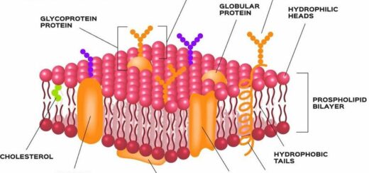
2. Cytoplasmic Membrane
It is a thin, elastic, semipermeable membrane consisting of a phospholipid bilayer.
Functions of the Cytoplasmic Membrane
- It is the osmotic barrier of the cell controlling the passage of nutrients into the cytoplasm and the end products of metabolism out of it.
- It is responsible for the electron-transport and oxidative phosphorylation because the cytochromes and other respiratory enzymes are located on it.
- It is responsible for the active transport of nutrients due to the presence of the permeases on it.
- It secretes hydrolytic enzymes which degrade large organic polymers into smaller molecules enough to penetrate the cytoplasmic membrane.
B. Cytoplasm and its components
1. Ribosomes
Ribonucleoprotein granules, visible by electron microscope and are sites of protein synthesis, they are composed of two subunits, 50 S and 30 S. Ribosomes are the sites of action of certain antibiotics.
2. Bacterial nucleoid
It consists of a single long double helix of DNA in the form of a closed circular thread about 1 mm long, devoid of the nuclear membrane and of the nucleolus.
3. Plasmid
Small circular DNA present as an extra-chromosomal element encodes a variable number of genes.
4. Inclusion granules
They represent excess metabolites stored as a nutrient reserve, e g lipid, glycogen, starch, sulphur or polyphosphates
C. External structure
1. Capsule
A capsule is a mucogelatinous layer surrounding the cell wall. It is not a constant structure and its presence is not linked with the viability of bacteria. The capsular substance is usually antigenic and is made of polysaccharides, though in some species, it is a protein in nature. In pathogenic bacteria, the capsule plays an important part in:
- Determination of virulence.
- Protecting bacteria against phagocytosis.
- Protecting the cell wall against attack by antibacterial agents.
2. Flagella
Long filamentous, wavy appendages, existing only in certain bacterial species, can be seen by electron microscope. The flagella are either displayed around the entire cell (peritrichous arrangement) or may be found at one or both poles (polar arrangement), Flagella are organs of locomotion that carry flagellar or H antigens made of a protein (flagellin).
3. Fimbriae or Pili
Pili are hair-like projections, found mainly in Gram-negative bacteria, They differ from flagella in being shorter, straight, and thinner. They are of two types:
- Common pili: They are structures by which bacteria can attach themselves to host cells (organs of adhesion), they are formed from an antigenic protein called pilin.
- Sex (F) pili. These pili are longer and thinner than common pili. (Refer to bacterial genetics).
You can subscribe to Science Online on YouTube from this link: Science Online
You can download Science online application on Google Play from this link: Science online Apps on Google Play
Features and classification of viruses, Defective viruses and Viral vectors used for gene therapy
Pathogenesis of bacterial infection, Classification of Pathogens & Bacterial virulence factors
Bacterial growth phases and Environmental factors required for bacterial growth