Liver function, site, shape, lopes, Biliary system, Gall bladder and Biliary ducts
The liver removes toxins from the body’s blood supply, It maintains healthy blood sugar levels, It regulates blood clotting, and performs hundreds of other vital functions, It is located beneath the rib cage in the right upper abdomen, It filters all of the blood in the body and breaks down poisonous substances, such as alcohol and drugs, It produces bile that helps digest fats and carries away waste.
Liver
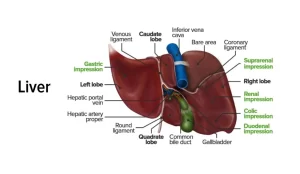
The liver lies under the diaphragm, in the right hypochondrium, epigastrium, and left hypochondrium. It is wedge-shaped, so it has five surfaces: superior, inferior, anterior, posterior, and right surfaces.
Lobes of the liver
It is divided into right large and small left lobes by:
- The attachment of falciform ligament on anterior and superior surfaces.
- Fissure for lig, venosum on the posterior surface.
- Fissure for lig. teres on the visceral surface.
It also contains caudate and quadrate lobes.
Relations of the liver
The diaphragm and base of the right lung and pleura are related to the superior, anterior, and right surfaces.
- The anterior surface is also related to ant. abdominal wall.
- Superior surface is also related to the heart and pericardium.
- Right surface is also related to the 7th to 11th ribs.
- Posterior surface: It is formed of a bare area, groove for IVC, caudate lobe (it has two processes: the caudate process anteriorly to the right, and the papillary process anteriorly to the left), fissure for ligamentum venosum and oesophageal notch.
Bare area of the liver: It is a triangular area related directly to the diaphragm (not covered by peritoneum), its base is formed by the groove for IVC, its apex is formed by the right triangular ligament, its superior and inferior boundaries are the two layers of the coronary ligament.
5- Inferior (Visceral) surface: it shows the following features and impressions:
- Gastric impression.
- Fissure for ligamentum teres.
- Quadrate lobe.
- Fossa for gall bladder.
- Duodenal impression.
- Renal impression.
- Supra renal impression.
- Colic impression.
- Tuber omental (elevated area in the left lobe related to the lesser omentum).
Quadrate lobe
It is a rectangular part of the inferior surface of the liver. It is bounded by:
- the inferior border of the liver anteriorly.
- porta hepatis posteriorly.
- gall bladder fossa on the right side.
- fissure for ligamentum teres on the left side.
It is related to the transverse colon (anteriorly), pylorus, and 1st part of the duodenum (middle) and lesser omentum (posteriorly).
Caudate lobe
It is related on the right side to the groove for the IVC. On the left side to a fissure for the ligamentum venosum superior to the ligamentum venosum as it curves to join the IVC and inferior to porta hepatis, The lower and right part of the caudate lobe forms a projection called the caudate process which forms the superior boundary of the epiploic foramen.
The lower and left part of the caudate lobe forms a projection called the papillary process, It is related posteriorly to the diaphragm, descending thoracic aorta, and T. 12.
Porta hepatis
It forms the hilum of the liver. Anteriorly it is bounded by the quadrate lobe and posteriorly by the caudate lobe and process, It gives attachment to the lesser omentum, Structures passing through it:
- Right and left branches of hepatic ducts: anterior in position.
- right and left branches of the hepatic artery: intermediate in position.
- right and left branches of portal vein posterior in position.
- Lymphatics.
Blood supply of liver
- It receives blood from two sources: The hepatic arteries which divide into right and left branches and the portal vein which divides into right and branches.
- The venous drainage is by three hepatic veins which terminate in the inferior vena cava (right, left, middle).
Lymphatic drainage of liver
The liver is drained by portal lymph nodes then into the coeliac lymph nodes except the bare area of the liver drains into subphrenic lymph nodes or posterior mediastinal lymph nodes.
Peritoneal connections
- Falciform ligament, Its free lower border contains the ligamentum teres (round ligament of the liver).
- Upper layer of the coronary ligament.
- Lower layer of the coronary ligament. Right triangular ligament.
- Left triangular ligament.
- Lesser omentum.
Embryonic remnants
- Ligamentum teres: It connects the umbilicus with the left branch of the portal vein. It represents the obliterated umbilical vein.
- Ligamentum venosum: It connects the left branch of the portal vein with the IVC. It represents the obliterated ductus venosus.
Areas of the liver not covered by the peritoneum
- Bare area.
- Groove for IVC.
- Porta hepatis.
- Fossa of gall bladder.
- Fissures for ligamentum teres and ligamentum venosum.
Hepatic segmentation
It depends on the vascular distribution to the liver (according to the venous drainage by the hepatic veins), It is divided into right and left lobes by an imaginary line passing through the IVC and the fossa of the gall bladder, This includes caudate and quadrate lobes as parts of the left lobe.
Surface anatomy of liver and gall bladder
A- Between 3 points:
- A point in the left 5th intercostal space 3 ½ inches from midline.
- A point in the right 5th rib midclavicular line.
- A point in the right 7th ribs in the midaxillary line.
B- Right surface: from right 7th-11th ribs (mid-axillary line).
C- Fundus of gall bladder: the tip of right 9th costal cartilage.
Biliary system
It consists of the gall bladder and the biliary ducts.
Gall bladder
It is a pear-shaped sac situated on the inferior surface of the liver. It has three parts; fundus (at the tip of the right ninth costal cartilage), body, and neck. The neck is continuous with the cystic duct and contains a valve. It is supplied by the cystic artery (from the right branch of the hepatic artery). Its venous drainage goes to the cystic vein which drains to the right branch of the portal vein.
Biliary ducts
1- Hepatic ducts:
- These are two ducts (right, and left), one from each lobe of the liver.
- They lie in front of the two branches of portal veins and the hepatic artery.
- They unite in the right part of the porta hepatis to form the common hepatic duct.
2- Common hepatic duct:
- It is one inch long. It descends in front of the portal vein and to the right of the hepatic artery. It joins the cystic duct to form the bile duct.
3- Cystic duct:
- It is one and a half inches long. It is S-shaped. It joins the common hepatic duct at an acute angle to form the bile duct just below the porta hepatis.
4- Common Bile duct:
It is 3 inches long, Its course is divided into 3 parts:
- It descends in the free margin of the lesser omentum anterior to the portal vein and on the right side of the hepatic artery.
- Then, it descends behind the 1st part of the duodenum and the portal vein is posterior to it.
- Then it passes behind the head of the pancreas where it joins the pancreatic duct to form the ampulla of Vater which opens in the posterior wall of the 2nd part of the duodenum below its middle, It forms an elevation in mucosa called. major duodenal papilla, The opening is guarded by the sphincter of Oddi.
You can download Science online application on Google Play from this link: Science online Apps on Google Play
Physiology & functions of Stomach, Composition of gastric secretion
Abdomen muscles, Blood Supply of Anterior Abdominal Wall & Rectus Sheath content
Stomach parts, function, curvatures, orifices, peritoneal connections & Venous drainage of Stomach


