Urinary system development, function and Congenital Malformations of the kidney
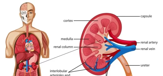
The urinary system removes the metabolic waste products such as uric acid, urea, and creatinine, It maintains electrolyte, water, and pH balance, It regulates blood pressure, blood volume and erythropoiesis, and vitamin D production, It consists of the kidneys, ureters, bladder, and urethra.
Development of the urinary system
The development of the urinary system is related to the development of the reproductive system; during the earlier stages.
Development of the kidney
Primordia: Intermediate mass of mesoderm: In the cervical part, it is segmented into groups of cells called nephrotomes. While in the caudal part, it gives a longitudinal ridge called nephrogenic, Three sets of kidneys appear following each other; Pronephros, Mesonephros & Metanephros.
1. Pronephros: Most primitive Kidney
The cervical nephrotomes give 5-7 pairs of pronephric tubules which open in a duct called the pronephric duct, The pronephros are non-functioning in mammals, The tubules degenerate by the 4th week, pronephric duct persists to become the mesonephric duct.

Development of the urinary system
2. Mesonephros
It is formed of about 80 mesonephric tubules. It differs from pronephros in the following:
- The mesonephric tubules are more caudal and more numerous (80).
- They have no special duct. They use the pronephric duct which is now called the mesonephric duct (Wolffian duct). The mesonephric duct elongates and opens into the cloaca.
Each mesonephric tubule has 2 ends; one end opens in the mesonephric duct while the other end differentiates into a cup-shaped Bowman’s capsule that is invaginated by capillaries forming the Glomerulus, Bowman’s capsule and Glomerulus make up the Renal Corpuscle, Mesonephric kidney functions for some time and then degenerates.
Some mesonephric tubules persist and give efferent ductules in males while in females, they give epophoron and, parophoron, The mesonephric duct forms the epididymis, vas deferens, and ejaculatory duct in males while it disappears in females leaving Gartner’s duct.
3. Metanephros
It is the permanent kidney. It begins functioning at 9 weeks, Urine is produced and released into the amniotic fluid. Primordia: It develops from two parts; the secretory part & the collecting part.
- The collecting part: The ureteric bud arises as an outgrowth from the lower end of the mesonephric duct near the cloaca, The ureteric bud elongates forming the ureter, renal pelvis, major calyces, minor calyces, and collecting ducts & tubules.
- The secretory part: The nephrogenic cord which is the caudal part of the intermediate-mass of mesoderm is divided into segments called metanephrogenic cap, Each segment lies opposite one collecting tubule.
The metanephrogenic cap forms the nephron:
- Glomerulus.
- Bowman’s capsule.
- Proximal and distal convoluted tubules.
- Loop of Henle.
The metanephrogenic cap forms a vesicle then a tubule that gets in contact with a collecting tubule, Contact with the collecting tubule is essential to produce proteases that break down the extracellular matrix between the metanephrogenic tubule and the collecting tubule, These tubules then become continuous.
Late Changes of kidney development
Branching of the calyces becomes larger, Kidneys ascend from the pelvic region to the abdominal region, The hilum of the kidneys was first anterior then with medial rotation 90 degrees, it becomes medial, Kidneys also undergo a lateral displacement that brings them in contact with the developing adrenal glands that fuse to their cranial pole.
Congenital Malformations of the kidney
- Renal agenesis means failure of the formation of one or the two kidneys, It may be isolated or associated with other congenital anomalies, It is always associated with oligohydraminos.
- Polycystic kidney means abnormal development of the collecting system, or failure of the collecting tubules and nephrons to join, The kidney contains many cysts. It may lead to renal failure, There are two forms of polycystic kidney namely the adult and the infantile forms.
- The pelvic kidney is due to failure of the kidney to ascend to the abdomen where it is kept in the pelvis.
- Horseshoe kidney: It occurs when the lower poles of both kidneys fuse together and this makes the kidneys fail to ascend.
Development of the ureter
Primordia: ureteric bud which is an outgrowth from the mesonephric duct, It develops from the caudal part of the mesonephric duct, Its elongates and its cranial end dilates to form the renal pelvis, major calyces, minor calyces, and collecting ducts & tubules, The developing bladder absorbs the lower part of the mesonephric duct till the origin of the ureters.
Congenital Malformation
Double ureter: Early splitting of ureter completely or partially; ureters open into the bladder separately, or unite and form one tube that opens in the bladder.
Development of the urinary bladder
The terminal part of the hindgut (cloaca), which is endodermal in origin, is divided by the cloacal septum into the anterior part, primitive urogenital sinus, and the posterior part that will form rectum and upper part of the anal canal.
Primordium: urogenital sinus, The urogenital sinus is divided into three parts.
- The cranial portion is continuous with the allantois and forms the bladder properly.
- The pelvic part of the sinus forms the prostatic urethra, the membranous urethra and bulbo urethral glands in the male and the membranous urethra and part of the vagina in females.
- The caudal portion, or definitive urogenital sinus, forms the penile urethra in males and the vestibule in females.
The caudal part of the mesonephric duct is absorbed into the posterior wall of the primitive urinary bladder till the origin of the ureters, This absorbed part will form the trigone of the bladder which is mesodermal in origin.
Development of the urethra
Primitive urogenital sinus gives primitive urinary bladder cranially and definitive urogenital sinus causally, The constriction in between gives the upper part of prostatic urethra in male and female urethra.
Definitive urogenital sinus
In males:
- The Cranial (pelvic) part gives the lower part of the prostatic urethra and membranous urethra.
- Caudal (phallic) part gives penile urethra except for the fossa navecularis (ectodermal in origin).
In females: It gives the membranous urethra and the vestibule of the vagina.
Congenital anomalies
- Urachal fistula: Caused by persisting allantois, Urine may drain from the umbilicus.
- Urachal cyst: Caused by failure of obliteration of the middle part of the allantois.
- Urachal sinus is caused by failure of the obliteration of the proximal or distal part of the allantois.
- Ectopia Vesicae (Extrophy of the bladder) is a rare and severe anomaly where both the anterior abdominal wall and the anterior wall of the bladder are absent and the posterior wall of the bladder (trigone and ureteric orifices) are exposed and protruded to the outside dribbling urine.
- Congenital recto-vesical fistula.
- Hypospadias: The urethra opens on the undersurface of the glans penis. might be perineal (scrotal), penile, or terminal, It is a common anomaly that might be an isolated defect or may be associated with other anomalies of the male genital system (as a part of ambiguous genitalia).
You can subscribe to science online on YouTube from this link: Science Online
You can download Science Online application on Google Play from this link: Science Online Apps on Google Play
Acid-base disturbances, metabolic acidosis causes, Effects of acidosis & alkalosis on the body
Physical properties of urine, Tests to evaluate kidney function & Normal constituents of urine
Histological structure of kidneys, Uriniferous tubules & Types of nephrons
Functions of Kidneys, Role of Kidney in glucose homeostasis, Lipid & protein metabolism