Placenta types, structure, function, development and abnormalities
The placenta offers oxygen & nutrients to the growing baby and it removes waste products from your baby’s blood. It develops in the uterus during pregnancy, It attaches to the wall of the uterus, and the baby’s umbilical cord arises from it. It is attached to the top, side, front, or back of the uterus.
The placenta
The placenta is the site of nutrient, gas exchange, and excretion between the fetus and mother. Placentas are a defining characteristic of placental mammals but they are found in marsupials and some non-mammals with varying levels of development.
Development of the placenta
At first, the chorionic villi cover the entire surface of the chorion. Later on, the villi opposite the decidua basalis continue to grow and expand to form chorion frondosum. While the villi related to the decidua capsularis degenerate and this part of chorion called chorion leave. The placenta is formed by:
- Mainly by the chorion frondosum (the fetal part, chorionic plate).
- A small extent by the decidua basalis (the maternal part, decidual plate).
Structure of the placenta
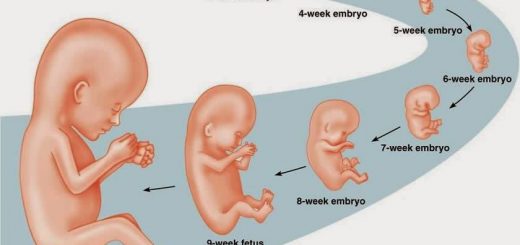
- Formation of the placenta started at the 4th month.
- The placenta has two components: the fetal part (chorion frondosum) and the maternal part (decidua basalis).
- Spaces between the villi appear and fuse together forming the intervillous spaces. They are filled with maternal blood. This blood leaked from the spiral arteries eroded by fetal chorionic villi.
- Later on, a number of decidual septae attached to the decidual plate are formed.
- These septa project into the intervillous spaces, but do not reach the chorionic plate, so it is an incomplete septum.
- As a result of these septa, the maternal surface of the placenta is divided into compartments; each one is called “cotyledon”.
- The intervillous spaces in all the cotyledons are communicating with each other. As pregnancy advances, the placenta increases in size by the formation of new villi, and in thickness due to the extensive arborization (branching) of the villi.
Gross appearance of full-Term Placenta
It is discoid shaped with a diameter of 15-25 cm, 3 cm thickness, and a weight of 500-600 gm (about 1/6 of the weight of a full-term fetus). It covers 15-30 % of the decidua. It is delivered approximately 30 minutes after the birth of the baby. It has two surfaces:
- Maternal surfaces are irregular, rough, reddish, and contains 15-20 cotyledons with deep grooves in between made by the decidual septa.
- The fetal surface is smooth and shiny (as it is covered by amnion). It has a number of chorionic umbilical vessels converging towards the umbilical cord, and the umbilical cord is attached centrally to this surface.
The placenta membrane (placental barrier)
It is the structures that separate the maternal and fetal blood. It is not a true barrier because few substances are able to cross it, most drugs in maternal blood can pass through it to the fetal circulation. Some of which can harm the fetus and cause major congenital anomalies. Early in pregnancy (till about 20-week gestation), the placental barrier is formed of four layers:
- The endothelial lining the fetal vessels.
- The connective tissue (primary mesoderm) of the villus.
- The cytotrophoblast layer.
- The syncytiotrophoblast.
After 20 weeks the cytotrophoblast degenerate so increases the permeability of the placenta, Towards the end of pregnancy, the fibrinoid material made of fibrin is formed on the surface of the villi to decrease the permeability, so the placental barrier is formed of this fibrinoid material, primary mesoderm, the syncytiotrophoblasts, and the endothelium of the fetal blood vessels.
The fetal circulation and maternal circulation are closed circulations, meaning that maternal blood and fetal blood do not mix.
Functions of the placenta
- Exchange of gases (respiration): Oxygen, and carbon dioxide are transported by simple diffusion.
- Exchange of nutrients, water, and electrolytes (nutrition): as amino acids, fatty acids, carbohydrates, and vitamins.
- Transmission of maternal antibodies to the fetus resulting in passive immunity.
- Excretion as fetal waste products e.g. urea and uric acid pass through it from fetal to maternal blood.
- Selective barrier (protection) against the transmission of diseases from the mother to the fetus. However many maternal infectious agents as viruses of rubella, measles, cytomegalovirus, and toxoplasma can cross the placenta causing severe congenital malformations or fetal death.
- Hormone production: By the end of the 4th month, the placenta secretes the following hormones by the syncytiotrophoblasts:
- Progesterone (it maintains the corpus luteum and preventing menses during pregnancy).
- Estrogenic (estriol) hormones.
- Gonadotrophins: as human chorionic gonadotropins (HCG), somato-mammotropin, human chorionic thyrotropin, and human chorionic corticotropin.
- Relaxin hormone to soften the ligaments of the pelvis in preparation for the birth of the fetus.
Abnormalities of the placenta
1. Shape abnormalities:
- Bilobed, trilobed, or horseshoe.
- Placenta membranacea (diffuse placenta), the thin layer of the placenta attaches to a large area of the uterus.
2. Number abnormalities:
- Double placentae.
- Triple placentae.
- Accessory placenta. It may cause severe postpartum hemorrhage if it is retained in the uterus after labor.
3. position abnormalities: Placenta previa where the placenta is attached to the lower uterine segment (due to low level of implantation of the blastocyst). It causes severe antepartum hemorrhage. There are three types of placenta previa:
- Placenta previa centralis: the center of the placenta covers the internal os of the cervix of the uterus.
- Placenta previa marginalis covers the internal os incompletely.
- Placenta previa parietalis is attached to the lower segment away from the internal os.
Placenta previa is usually diagnosed by ultrasonography during pregnancy and delivery must be by Cesarean section to avoid severe antepartum hemorrhage.
4. Abnormal penetration to the uterine wall:
- Placenta accreta: due to abnormal adhesion between the chorionic villi and the uterine wall due to excessive penetration of the endometrium.
- Placenta percreta: The chorionic villi penetrate the myometrium all the way to the perimetrium (uterine peritoneal covering).
In these two abnormalities, the placenta fails to separate from the uterus after the birth of the fetus and may cause severe postpartum hemorrhage.
5. Abnormalities due to the attachment of the umbilical cord:
- Battledore placenta: The cord is attached to the margin of the placenta. It is named so because of its resemblance to the bat used in the medieval game of battledore and shuttlecock.
- Velamentous placenta: The cord is not attached to the placenta, and the umbilical blood vessels reach the placenta by passing in the amniotic membrane.
Fetal membrane layers, Chorion, Amnion, Yolk sac & umbilical cord
Productive organs, Germ cells, Fertilization, Stages of cleavage, Implantation & Decidua
Second & third week of Embryonic development, Chorion & types of Chorionic villi
Third to eight weeks (Embryonic period), Embryo Folding & Results of folding
Placenta importance in fetal development & the most common placental problems