Second & third week of Embryonic development, Chorion & types of Chorionic villi
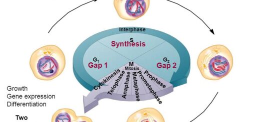
The embryo is the early stage of the development of the multicellular organism. Each embryo starts development as the zygote, a single cell resulting from the fusion of gametes, The zygote undergoes many rapid cell divisions, called cleavage, to form the blastula, then, the cells in a blastula-stage embryo start rearranging themselves into layers in a process called gastrulation.
Embryo
These layers will each give rise to different parts of the developing multicellular organism, such as the nervous system, connective tissue & organs. A newly developing human is referred to as the embryo until the ninth week after conception when it is then referred to as the fetus.
Second-week development (The Bilaminar Germ Disc)
Changes occurring during the second week of development:
1. Further differentiation of the Trophoblast
The trophoblast is formed of two-layer:
- An inner layer of mononucleated cells called the cytotrophoblast.
- An outer layer of syncytiotrophoblast, it is formed by migration of cytotrophoblasts after losing their cell membrane.
Syncytiotrophoblast will form finger-like processes called villi, they erode the material uterine tissues and vessels, leads to the formation of spaces between them filled with material blood, these spaces are called lacuna, The lacuna coaleases together to form intervillous space.
2. Formation of the bilaminar germ disc
Cells of the inner cell mass differentiate into two layers:
- A layer of small, cuboidal cells adjacent to the blastocyst cavity called the hypoblast (endodermal) layer.
- A layer of tall columnar epithelium adjacent to the amniotic cavity, called the epiblast (ectodermal) layer.
The cells of these two germ layers form the bilaminar germ disc (embryo).
3. The formation of the amniotic and yolk sac cavities
- Some vacuoles appear within the epiblast layer (ectodermal layer). They collect together to form a small cavity called the amniotic cavity.
- The cells of the epiblast (ectoderm) adjacent to the cytotrophoblast are called the amnioblasts.
- The amnioblasts form the roof of the amniotic cavity and the ectodermal layer of the embryonic disc forms the floor of the cavity.
- Another cavity is formed in the ventral aspect of the embryonic disc, called the primitive yolk sac (the exo-coelomic cavity). Its roof is formed of the endodermal layer of the embryonic disc.
4. Formation of the extra-embryonic mesoderm and extraembryonic coelom
The endoderm of the wall of the yolk sac gives new cells called the extra-embryonic (primary) mesoderm which surrounds the amnion, and yolk sac. A cavity appears in the extra-embryonic mesoderm called the extra-embryonic coelom (the chorionic cavity). The extraembryonic primary mesoderm is divided into two layers by the extra-embryonic coelom:
- An outer layer lining the trophoblasts and an inner layer covers the amnion, called the extra-embryonic somatopleuric mesoderm. The two layers are connected by a body stalk.
- A layer covering the yolk sac called the extra-embryonic splanchnopleuric mesoderm. The intraembryonic coelom is a space that appears in the intraembryonic mesoderm, it is in the shape of an inverted U.
It will give rise to:
- The future pericardial cavity. at the center of the coelom.
- The future pleural cavity. on either side of the pericardial cavity.
- The future peritoneal cavity. lateral to the pleural cavities on either side and connected to the extraembryonic coelom.
Clinical application
Syncytiotrophoblast secretes human chorionic gonadotrophin hormone which prevents the degeneration of the corpus luteum. It also stimulates the production of progesterone from the ovary. By the end of the 2nd week, the amount of this hormone will be sufficient to be detected in the maternal blood and urine. This is the basis of the pregnancy test.
The second week of development is the week of twos, because of the following:
- Trophoblast differentiates into 2 layers, cytotrophoblast & syncytiotrophoblast.
- Inner cell mass differentiates into 2 layers, epiblast & hypoblast.
- Primary mesoderm splits into somatopleuric primary mesoderm & splanchnopleuric primary mesoderm.
- Starting with the formation of the amniotic and yolk sac cavity.
The chorion
Structure of the chorion: The chorion is formed of:
- An outer layer consisting of the two layers of trophoblast.
- An inner layer of primary mesoderm (extra-embryonic mesoderm).
The Chorionic villi
Types of the villi according to structure:
- Primary villi is the early strand of trophoblasts. It is formed of an inner cytotrophoblast and an outer syncytiotrophoblast. The blood spaces in between the villi called the intervillous spaces filled with maternal blood.
- Secondary villi: The extra-embryonic mesoderm cells invade the primary villi forming a core inside them.
- Tertiary villi: umbilical blood vessels are formed in the mesodermal core of the secondary villi.
Types of villi according to their function:
- 1. Anchoring villi: They fix the embryonic, so, they penetrate deeply the decidua basalis of the uterus.
- 2. Free (Nourishing) villi: They are free and do not reach the decidua. They extend into the maternal blood of the intervillous spaces for the nutrition of the embryo.

Second & third week of embryonic development
Third week of development (Trilaminar germ disc)
Gastrulation (Formation of three germ layers embryo)
- Gastrulation is the most characteristic event occurring during the 3rd weak. During this period, the embryo is referred to as gastrula.
- It is the process by which the bilaminar embryonic disc is converted into a trilaminar embryonic disc.
- Gastrulation begins with the formation of a midline groove on the surface of the caudal part of the epiblast of the bilaminar embryonic disc it is called a primitive steak.
- The cephalic end of this steak is called a primitive node. A groove appears at the center of the node called a primitive pit.
Formation of the primitive streak (groove)
Cells of the ectoderm (epiblast) migrate in the direction of the primitive streak, they slip to under the epiblastic layer through the primitive streak to form the endodermal layer of the embryonic disc. Some of the invaginated epiblastic cells form the intraembryonic mesoderm, thus, the ectoderm or the epiblast is the source of the three germ layers of the embryo).
The intraembryonic mesoderm migrates in all directions forming a complete layer of cells separating the ectoderm dorsally from the endoderm ventrally (trilaminar embryo).
Formation of the notochord
Some mesenchymal cells migrate cranially from the primitive node and pit (deep to epiblast), forming a median cellular cord; the notochord.
Functions of the notochord
- It forms the axis around which the axial skeleton develops (bones of the head and vertebral column).
- It stimulates the overlying ectoderm to form the central nervous system.
- The notochord degenerates and disappears as the bodies of the vertebrae form. Its remnant is the nucleus pulposus of the intervertebral discs found in between the vertebrae.
Clinical application
The third week of development is a very sensitive period in embryonic development. Many factors such as drugs, alcohol, or irradiation to the mother may cause congenital anomalies to her embryo.
At the end of the fourth week, the primitive streak shows regressive changes, rapidly shrinks, and soon disappears. Sometimes, remnants of the primitive streak persist in the sacrococcygeal region. These pluripotent cells proliferate and form tumors known as sacrococcygeal teratomas.
Reproduction in Humans, Structure of Male & Female reproductive (genital) system
Productive organs, Germ cells, Fertilization, Stages of cleavage, Implantation & Decidua
Third to eight weeks (Embryonic period), Embryo Folding & Results of folding
Fetal membrane layers, Chorion, Amnion, Yolk sac & umbilical cord
Placenta types, structure, function, development & abnormalities