Excitability changes in skeletal muscle fibers during activity and Causes of muscle fatigue
Muscle fatigue decreases your muscles’ ability to perform over time, It can occur anywhere on the body, It is associated with exhaustion state, often following strenuous activity or exercise, if you experience fatigue, the force behind your muscles’ movements decreases, causing you to feel weaker. Exercise is a popular cause of muscle fatigue, this symptom can be the result of other health conditions, too.
Excitability changes in skeletal muscle fibers during activity
Generally speaking the excitability changes in skeletal muscle fiber is the same as in the nerve fiber.
Mechanical changes in skeletal muscle fibers during activity
In relation to muscle, the electrical response is followed by the mechanical response (muscle contraction). (Molecular basis of muscular contraction).
Sliding filament theory (the molecular basis of muscular contraction)
Shortening or contraction of muscle occurs by sliding the thin filament of the myofibrils over the thick filament. During rest i.e. relaxation of the muscle, Troponin T is tightly bound to tropomyosin forming a troponin-tropomyosin complex. This complex covers the binding sits for myosin heads on the actin filament, so contraction is inhibited.
Once troponin C is combined with calcium ions, the action of the troponin-tropomyosin complex disappears, tropomyosin moves laterally leading to uncovering of the binding sites of myosin heads on the actin filament, and the process of contraction starts.
The heads of myosin link to actin at a right angle, then swiveling of the myosin heads followed by detaching and reattachment to the next linking site. The energy for muscle contraction is derived from the breakdown of ATP. The heads of myosin molecules have ATPase activity.
Excitation-contraction coupling mechanism
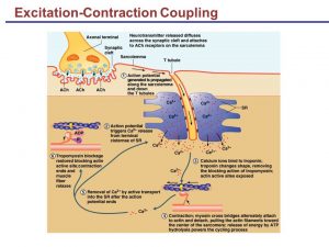
The excitation-contraction coupling mechanism is the process by which an action potential initiates the muscle contraction. It involves the following steps:
- Stimulation of myelinated motor nerve supplying a skeletal muscle leading to a generation of the action potential, which spreads along both sides of the sarcolemmal membrane.
- The action potential spreads to the depth of the myofibrils via the T-tubular system (extending from the sarcolemmal membrane).
- Release of calcium ions from the lateral sacs of the sarcoplasmic reticulum and its diffusion to the thick and thin filaments.
- The binding of Ca++ to troponin C, uncover the binding sites of myosin on the actin filaments. This is due to inhibition of the troponin-tropomyosin complex and lateral movement of tropomyosin. ATP is splitted to supply the energy needed for muscular contraction.
- Sliding of the thin filaments over the thick filaments, due to the formation of cross-linkages between myosin and actin, leading to muscular contraction.
- More and more shortening is obtained by disconnection, cocking (return to a right angle), reconnection and, swiveling myosin heads to the binding sites on actin filaments.
- Active reuptake of calcium by the sarcoplasmic reticulum to be stored for the next action potential. The energy needed is obtained from the breakdown of ATP, so ATP is used in muscular contraction and relaxation.
- Once the calcium is reuptaken by the sarcoplasmic reticulum, the interaction between myosin and actin stops, and muscular relaxation occurs.
Types of muscular contraction
There are two types of muscle contraction:
1- Isotonic contraction
Isotonic contraction occurs when the muscle contracts against a light or moderate load, this contraction leads to shortening of the muscle and movement of the load. One-fourth of the energy of contraction is converted into beneficial work, and the remaining 75% is dissipated as heat to the atmosphere.
The work done by the muscle = Weight (in kg) x height or distance moved by the weight (m) = kg/m.
If either the weight or height is zero no work is done.
2- Isometric contraction
This type of contraction occurs when the muscle contracts against a heavy load. The muscle does not shorten; its length remains the same (isometric), and the load does not move, therefore the muscle does not perform any work. The tension in the muscle increases very much during the contraction. Isometric contraction is observed when muscle contracts to move a weight that is too heavy to be moved. All the energy will be converted into heat.
Muscle fatigue
This is a temporary decrease in the work capacity of skeletal muscles following prolonged or forceful contraction, Muscle fatigue is the defense mechanism that protects the muscle from reaching the point at which it can no longer produce ATP.
Causes of muscle fatigue
- Depletion of energy-producing substances (glycogen, ATP, CP).
- The local increase in ADP and inorganic phosphate from ATP breakdown may directly interfere with cross-bridge cycling and/or block calcium release and uptake by the sarcoplasmic reticulum.
- Accumulation of lactate may inhibit key enzymes in the energy-producing pathways and/or excitation-contraction coupling.
- Reduction to a small extent in nerve signal transmission at the neuromuscular junction after intense prolonged muscular activity.
- Interruption of blood flow through a contracting muscle leads to almost complete muscle fatigue within 1-2 min because of loss of nutrient supply, especially O2.
Neuro-Muscular Junction properties, Functions and types of skeletal muscles
Muscles types, Skeletal muscle parts, Lymphatic system structure and function
Muscular system, Structure of skeletal muscle, Muscles properties & functions
Muscular tissue types, function, structure, definition & anatomy
Medial compartment of thigh muscles, Growth & regeneration of smooth muscle fibers


