Function of sensory nervous system, Histological structure of ganglia and receptors
The sensory nervous system is responsible for processing sensory information, It is a part of the nervous system, It is composed of sensory receptors, neural pathways, and parts of the brain involved in sensory perception, the receptive fields have been identified for the visual system, auditory system, and somatosensory system, so far.
Histological structure of ganglia and receptors
They are collections of nerve cell bodies, covered by a connective tissue capsule, and lie outside the CNS in the pathway of nerves. There are two types of ganglia:
- Cranio-spinal ganglia (sensory ganglia).
- Autonomic ganglia: either sympathetic or parasympathetic ganglia.
Histological organization of cranio-spinal ganglia
- Site: they are associated with the dorsal root of all spinal nerves (dorsal root ganglia) and the sensory root of some cranial nerves e.g. trigeminal nerve.
- Morphology: larger than autonomic ganglia and oval in shape.
- Connective tissue capsule & septa: thick capsule with thick septa running parallel to the capsule.
- Ganglion cells: large pseudounipolar neurons with central nuclei. The ganglion cells are arranged in a few groups in the substance of the ganglion. Each ganglion cell is surrounded by a complete capsule of satellite cells (neuroglia cells).
- Nerve fibers: thick, myelinated running parallel to the capsule. They represent the central and peripheral processes of the ganglion cells.
- Synapse: not present.
- Function: sensory.
Histological organization of autonomic ganglia
- Site: they are located along the sympathetic chain and the parasympathetic nerves.
- Morphology: smaller than cranio-spinal ganglia and rounded in shape.
- Connective tissue capsule & septa: thin capsule with thin septa running irregularly in the ganglion.
- Ganglion cells: smaller stellate multipolar neurons with eccentric nuclei. The ganglion cells art numerous and scattered in the substance of ganglion. Each ganglion cell is surrounded by an incomplete capsule of satellite cells.
- Nerve fibers: thin mostly unmyelinated & irregularly distributed. They represent the pre-& post-ganglionic nerve fibers.
- Synapse: present between the preganglionic fibers and the ganglion cells.
- Function: visceral motor.
Histological organization of sensory nerve endings

Sensory nervous system
Nerve endings are biological transmitters which are classified as follows:
- Motor nerve endings (effectors): they are terminals of axons that transmit impulses from the CNS to the muscle fibers to contract (motor end plate) or to glandular cells to secrete.
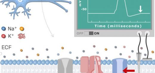
- Sensory nerve endings (receptors): they receive various forms of physical and chemical stimuli. Following their stimulation, the resulting receptor potentials are transmitted as sensory input to the CNS.
According to their histological structure, receptors can be classified into:
A. Non-encapsulated receptors: they are not covered by a connective tissue capsule. These include:
1. Free nerve endings: these are naked nerve fibers (unmyelinated & without neurilemma). These include:
a. Pain receptors (nociceptors):
Site:
- In the epithelium of the skin, corneal and buccal mucosa. They penetrate the basement membrane and branch freely in-between the epithelial cells. In the skin, they reach up to the granular layer of the epidermis.
- In the connective tissue of the tympanic membrane and dental pulp.
- Nociceptors are also found in the muscle, joints, bone, and viscera.
b. Thermoreceptors:
Site: they ramify in the papillary layer of the skin and run at dermo-epidermal junction parallel to the skin surface.
2. Plexus of Bounet (Peritrichial free nerve endings):
- Site: they ramify around the hair follicles.
- Function: they are receptors for touch, stimulated by hair movement.
3. Merkel’s discs: touch receptor.
B. Encapsulated receptors: they are more specialized and exhibit a characteristic CT capsule. These include:
1. Meissner’s corpuscles
- Site: they are present in the dermal papillae in the skin and more in the palmar surface of the tips of fingers, the skin of lips, and nipples.
- Shape: small encapsulated oval structure lying with its long axis perpendicular to the surface of the skin.
- Histological structure: the corpuscle consists of flattened Schwann cells that are transversely arranged within the corpuscle.
- Innervation: the sensory nerve fiber loses its myelin and neurilemmal sheaths, enters the corpuscle from its lower pole, and runs a spiral course giving branches between the cells of the corpuscle.
- Function: touch receptor.
2. Pacinian corpuscles (Corpuscles of Pacini)
Site:
- Lie deep in the dermis and hypodermis of the skin; numerous in the fingers, external genitalia, and breast.
- They are also present in the skeletal muscles, tendons, and joint capsules.
- In the CT of the viscera (pancreas and urinary bladder).
Shape: large ovoid lamellated corpuscle.
Histological structure: the core of the corpuscle is formed of 30-60 concentric layers of flattened Schwann cells and fibroblasts. These layers are separated by intercellular spaces containing collagen fibers and tissue fluid. So in the histological section, it appears like a sliced onion.
Innervation: the nerve fiber loses its sheaths on entering the corpuscle, runs in the long axis through the central core to end at the opposite pole of the corpuscle.
Function: pressure and vibration receptor.
3. Ruffini’s corpuscles
- Site: lie deep in the dermis, oriented parallel to the skin surface (more in the soles).
- Histological structure: Ruffini’s corpuscle is a small elongated structure containing collagen fibers and fibroblasts and is surrounded by a thin CT capsule.
- Innervation: the nerve fiber enters from the side of the corpuscle, loses its myelin, and branches repeatedly between the collagen fibers. It is stimulated by displacement of the collagen fibers.
- Function: pressure receptor.
- Innervation: the nerve fiber enters from the side of the corpuscle, loses its myelin, and branches repeatedly between the collagen fibers. It is stimulated by displacement of the collagen fibers.
- Function: pressure receptor.
4. Muscle spindles
Site: they are distributed between the skeletal muscle fibers mainly near the musculo-tendinous junctions.
Histological structure: the spindle is an encapsulated fusiform structure situated parallel to the striated muscle fibers and attached to them by its ends, So, when the muscle is stretched, the spindle is stimulated, The muscle spindle is formed of 2 types of small, modified muscle fibers called the intrafusal muscle fibers (to distinguish them from the ordinary extrafusal skeletal muscle fibers), The intrafusal fibers are present in the interior of the spindle’s cavity that is filled with a gelatinous substance.
Types & innervation of intrafusal muscle fibers:
- Nuclear bag fibers: they are 2-4 in number. Each has a central non-contractile region expanded like a bag and contains many aggregated nuclei.
- Nuclear chain fibers: they are 6-8 in number and they are shorter and narrower than nuclear bag fibers. Each has a central non-contractile region not expanded and contains nuclei arranged in the form of a chain.
Innervation: both afferent (sensory) and efferent (motor) nerve fibers pierce the CT capsule and supply the intrafusal muscle fibers. They include:
- Primary afferent fibers (annulospiral endings) are formed of thick axons which encircle the nuclear region of the 2 types of intrafusal fibers.
- Secondary afferent fibers (flower spray endings) are formed of smaller axons which end at the peripheral part of the nuclear region of nuclear chain fibers only.
- Motor nerves (efferent fibers) formed of fine myelinated gamma efferent fibers arising from small gamma motor neurons of the anterior horn of the spinal cord. They innervate the peripheral contractile regions of the intrafusal fibers and end in motor endplates. Their principal role is to maintain the sensitivity of the muscle spindle.
Function: receptor for proprioceptive sensation specialized for monitoring changes in length and the degree of stretch of the muscle stretch reflex receptor
5. Tendon spindle “Golgi tendon organ”
- Site: in the tendons near the musculo-tendinous junction.
- Histological structure: The tendon spindle is smaller than muscle spindles and contains a group of wavy collagen fibers instead of the intrafusal muscle fibers.
- Innervation: the sensory nerve fiber loses its myelin, enters the spindle and gives multiple branches that ramify between the collagen fibers. No motor nerve endings are present.
- Function: receptor for proprioceptive sensation. It is stimulated by the straightening of collagen fibers of muscle tendon. It is specialized for monitoring the force (tension) produced by muscle contraction.
You can subscribe to science online on YouTube from this link: Science Online
You can download Science Online application on Google Play from this link: Science Online Apps on Google Play
Physiology of sensory receptors, Coding of sensory information and Somatic sensation
Development and Abnormalities of the face, palate, tongue and Nerve supply of face processes
Cranial nerves types, Facial, Vestibulocochlear, Glossopharyngeal, Vagus, Accessory & Hypoglossal
Triangles of the neck contents, Structure of Anterior and Posterior triangle of the neck
Facial muscles function, anatomy, arteries, veins, names & expressions
Skull function, anatomy, structure, views & Criteria of neonatal skull