Physiology of sensory receptors, Coding of sensory information and Somatic sensation
Sensory receptors are specialized structures that act as both: Detectors and Transducers. Detectors detect changes in the external or internal environment of the body. Transducers convert the energy of the change they have detected into electrical impulses.
Physiology of sensory receptors
According to the type of stimulus, receptors can be classified into:
Mechanoreceptors: respond to mechanical stimuli and include:
- Touch, pressure, and vibration receptors are located in the skin.
- Proprioceptors are located in muscles, tendons, and joints.
- Baroreceptors are located in the aortic arch and carotid sinus.
- Stretch receptors are located in the alveoli of the lungs and right atrium.
Thermoreceptors: stimulated by the thermal form of energy and detect changes in temperature and include.
- Cold receptors.
- Warmth receptors.
Chemoreceptors are stimulated by the chemical forms of energy: include:
- Taste receptors.
- Olfactory receptors.
- Glucoreceptors and osmoreceptors in the hypothalamus.
Nociceptors: stimulated by any form of energy potent enough to cause tissue damage.
Electromagnetic receptors: respond to the electromagnetic waves of light. Include the rods and cones in the retina of the eye.
Properties of receptors
A. Differential sensitivity (specificity)
Each receptor is most sensitive to one particular type of stimulus called its adequate stimulus. The sensory fiber from the receptor terminates in an area of the brain that gives the sensation perceived by the receptor. The sensation perceived as a result of stimulation of a receptor is called the modality of sensation.
Some receptors can be stimulated by stimuli other than their adequate stimulus provided the intensity of stimulation is sufficiently high, but will still be giving its own specific modality of sensation.
Differential sensitivity of receptors is illustrated by Muller’s law of specific nerve energies which states that each receptor is most sensitive to one specific type of stimulus called its adequate stimulus giving rise to one type of sensation regardless of the method of stimulation because the sensation perceived depends on the area in the brain ultimately activated.
B. Excitability
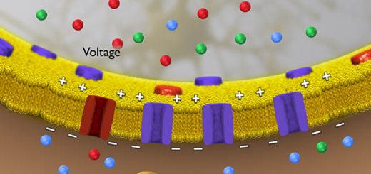
This is the ability of the receptor to respond to stimuli. In the resting condition, the receptor is in a polarized state i.e. its outer surface is positively charged while its inner surface is negatively charged with a resting membrane potential of about 70-mV.
On stimulation, the inflow of Na+ ions occurs as a result of the opening of Na+ channels in the receptor membrane producing a local, graded potential called the receptor or generator potential, When the receptor potential reaches the threshold value, the action potentials are generated in the first node of Ranvier of the afferent sensory fiber. An increase in the amplitude of the receptor potential produced by increased stimulus intensity causes an increase in the action potential frequency in the afferent sensory fiber.
Properties of the receptor potential
It is a local, unpropagated potential whereas action potentials are propagated. It is a graded potential i.e. It does not obey the all or none rule. It has no refractory period, so it can be summated. The receptor potential is caused by activation of mechanically gated channels whereas action potentials are caused by activation of voltage-gated channels. It is not blocked by local anesthetics, whereas action potentials are blocked by these drugs.
C. Adaptation:
Definition: this is the decrease in the number of nerve impulses in the sensory fiber due to a decline in the amplitude of the receptor potential by a constant maintained stimulus.
Types: according to the rate of adaptation, receptors are divided into:
- Rapidly adapting (phasic) receptors: as touch and olfactory receptors.
- Slowly adapting (tonic) receptors: as pain, baroreceptors, and muscle stretch receptors.
Mechanisms of adaptation
Mechanisms of adaptation may be due to one or more of the following mechanisms:
- Loss of the stimulus energy in the surrounding tissues by constant stimulation.
- The gradual decrease of the excitability of the first node of Ranvier.
- Accommodation of the afferent nerve fiber to the generator potential (receptor potential). This probably results from the inactivation of the sodium channels in the nerve fiber membrane.
Coding of sensory information
This is the ability of the brain to discriminate the modality, locality, and intensity of different stimuli although all sensations reach the brain in the form of nerve impulses.
- Modality discrimination: It depends on the differential sensitivity of the receptors. Discrimination of the modality of a sensation by the brain depends on the adequate stimulus to which the receptor is specialized and on the area of the brain ultimately activated.
- Locality discrimination: It depends on the fact that each receptor has a specific pathway to the sensory cortex where different parts of the body are represented. Therefore, stimulation of a sensory pathway anywhere along its course to the sensory cortex produces sensation referred to the location of the receptor.
- Intensity discrimination: It depends on the frequency of discharge of action potentials (nerve impulses) in the sensory fiber. A high frequency of discharge is interpreted by the sensory cortex as an increased intensity of stimulation. Discrimination of intensity also depends on the number of receptors stimulated since stronger stimuli tend to stimulate a greater number of receptors.
Somatic sensation
These are sensations from the skin and deeper structures (muscles, bones, tendons, and joints). According to their nature, somatic sensations are classified into:
A. Mechanoreceptive sensations: resulting from stimulation of mechanoreceptors include:
- Tactile sensations: touch sensation, pressure sense, vibration sense
- Proprioceptive (kinesthetic) sensations: subdivided into:
- Conscious proprioceptive sensations: senses of position and movement of joints.
- Unconscious proprioceptive sensations: senses of muscle length and tension.
B. Thermoreceptive sensations: resulting from stimulation of thermoreceptors and include:
- Cold sensation
- Warmth sensation
C. Pain sensation: resulting from stimulation of nociceptors. Itch sensations are produced by stimulation of pain receptors due to tissue irritation for example by an insect bite.
A. Mechanoreceptive sensations
They include:
1. Tactile mechanoreceptive sensations:
I. Touch sensation:
- Light touch: it is a touch sensation that is not sharply localized and results from the stimulation of tactile receptors in the superficial layers of the skin. It is tested using a piece of cotton wool on different dermatomes.
- Tactile discrimination (2-point discrimination): it is the ability to perceive, with closed eyes, two touch stimuli applied to the skin at the same time as two separate points of touch provided that the distance between the two points is more than a certain threshold variable in different areas of the body.
2. Pressure sensation: it is the ability to discriminate different weights and generally results from stimulation of tactile receptors in deeper layers of the skin.
3. Vibration sense: it results from rapidly repetitive sensory signals from tactile receptors.
4. Proprioceptive (kinesthetic) sensations:
- Conscious proprioception: includes the sense of joint position and the sense of joint movement, The receptors located in the joints provide the sensory information to the cerebral cortex which in turn, uses this information to generate conscious awareness of the orientation of different parts of the body to each other.
- Unconscious proprioception: includes the senses of muscle length and tension. The impulses arising from the proprioceptors mediating this type of sensation are relayed to the cerebellum and serve information about the momentary state of muscle contraction, degree of tension and length of muscles.
B. Thermoreceptive sensations
Cutaneous thermoreceptors are specialized free nerve endings, found with the highest density in the skin of the hands and face. Cutaneous thermoreceptors monitor skin temperature, not body temperature, and are thus useful in detecting changes in external environmental temperature, This information reaches the hypothalamus, which is the heat-regulating center to initiate nervous and humoral mechanisms for maintaining constant body temperature despite changes in environmental temperature.
Cutaneous thermoreceptors are innervated by small myelinated Aδ and unmyelinated C fibers that travel to the brain together with pain sensation.
Thermoreceptors include:
- Cold receptors: respond to temperatures between 10° and 35° C and are normally silent above 40° C, however, they may give a brisk discharge at 45° C producing a false cold sensation called paradoxical cold.
- Warmth receptors: respond to temperatures between 35° C to 45° C. Temperatures between 0° C and 10° C or above 45° C stimulate thermosensitive pain receptors namely cold-pain and hot-pain receptors respectively. At 0° C, all receptors stop discharging, a fact made use of cold for local anaesthesia.
You can subscribe to science online on YouTube from this link: Science Online
You can download the application on Google Play from this link: Science Online Apps on Google Play
Function of sensory nervous system, Histological structure of ganglia and receptors
Development and Abnormalities of the face, palate, tongue and Nerve supply of face processes
Lymphatics of head and neck, superficial vessels, deep vessels & Abnormalities of branchial arches
Cranial nerves types, Facial, Vestibulocochlear, Glossopharyngeal, Vagus, Accessory & Hypoglossal
Facial muscles function, anatomy, arteries, veins, names & expressions
Skull function, anatomy, structure, views & Criteria of neonatal skull


