The Ultimate Guide to the Esophagus: 12 Powerful Facts About Structure, Function & Common Disorders
The esophagus is a muscular tube that plays a vital role in the digestive system by transporting food and liquids from the mouth to the stomach. Although it appears simple, the esophagus has a complex structure and performs highly coordinated movements that are essential for swallowing and digestion. Disorders affecting the esophagus can cause pain, difficulty swallowing, acid reflux, and serious health complications if left untreated.
Esophagus
The esophagus is a muscular tube that connects the throat (pharynx) to the stomach, allowing food and liquids to pass through during swallowing. It is part of the digestive system and is about 8 to 10 inches (20-25 cm) long in adults. Understanding the anatomy and function of the esophagus helps people recognize early warning signs of esophageal diseases and seek proper medical care before complications develop.
Structure and Anatomy of the Esophagus
The esophagus is approximately 25 centimeters long in adults and connects the throat (pharynx) to the stomach. It is located behind the trachea and passes through the chest cavity before entering the abdomen. The wall of the esophagus contains several layers that help move food efficiently toward the stomach.
The inner lining, known as the mucosa, protects the esophagus from friction and damage caused by swallowed food. Beneath this layer are muscles that contract rhythmically during swallowing, a process called peristalsis. These muscular contractions push food downward smoothly and prevent it from moving backward.
The esophagus also contains two important sphincters. The upper esophageal sphincter controls the entry of food into the esophagus, while the lower esophageal sphincter prevents stomach acid from flowing back upward into the esophagus. Weakness in the lower sphincter is one of the main causes of acid reflux disease.

Esophagus parts
Structure of the Esophagus
Layers of the Esophageal Wall:
- Mucosa: The innermost layer, which produces mucus to help food pass smoothly.
- Submucosa: Contains glands and blood vessels.
- Muscularis: Muscle layer that contracts in waves (peristalsis) to push food downward.
- Adventitia: The outer layer, which connects the esophagus to nearby structures.
Sphincters
- Upper Esophageal Sphincter (UES): Prevents air from entering the esophagus.
- Lower Esophageal Sphincter (LES): Prevents stomach acid from flowing back into the esophagus (acid reflux/GERD).
Function of the Esophagus
- Moves food from the mouth to the stomach through peristalsis (wave-like muscle contractions).
- Prevents acid reflux with the LES.
- Protects itself from damage using mucus and constant cell renewal.
The primary function of the esophagus is to transport food, liquids, and saliva from the mouth to the stomach. During swallowing, the muscles of the esophagus contract in a coordinated sequence that safely moves food through the digestive tract.
The esophagus also helps protect the airway during swallowing. Special reflexes close the airway temporarily so that food does not enter the lungs. This process is essential for safe eating and breathing coordination.
Another important function is preventing acid reflux. The lower esophageal sphincter acts like a valve that keeps stomach acid inside the stomach. When this sphincter becomes weak or relaxes abnormally, acidic contents can irritate the esophagus and cause heartburn or inflammation.
Common Esophageal Disorders
- Gastroesophageal Reflux Disease (GERD): Acid from the stomach flows back into the esophagus, causing heartburn.
- Esophagitis: Inflammation of the esophagus due to infection, acid reflux, or allergens.
- Esophageal Stricture: Narrowing of the esophagus, often due to scarring from acid reflux.
- Barrett’s Esophagus: A precancerous condition caused by long-term acid reflux.
- Esophageal Cancer: Often linked to smoking, alcohol, or chronic acid reflux.
Several medical conditions can affect the esophagus and interfere with swallowing or digestion. Gastroesophageal reflux disease (GERD) is one of the most common disorders and occurs when stomach acid repeatedly flows back into the esophagus. This condition may cause heartburn, chest discomfort, and chronic irritation.
Esophagitis refers to inflammation of the esophagus and may result from acid reflux, infections, allergies, or certain medications. Symptoms often include pain during swallowing and throat discomfort.
Another serious disorder is achalasia, a condition in which the lower esophageal sphincter fails to relax properly, making it difficult for food to enter the stomach. Patients may experience difficulty swallowing, regurgitation, and weight loss.
Esophageal cancer is a potentially life-threatening disease that may develop due to chronic acid reflux, smoking, alcohol use, or Barrett’s esophagus. Early diagnosis significantly improves treatment outcomes.
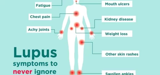
Symptoms of Esophageal Problems
Symptoms of esophageal disorders can vary depending on the condition, but commonly include:
- Difficulty swallowing.
- Heartburn.
- Chest pain.
- Regurgitation of food or acid.
- Chronic cough.
- Hoarseness.
- Unexplained weight loss.
- Painful swallowing.
Persistent symptoms should never be ignored because they may indicate a serious underlying disease.
Diagnosis and Treatment
Doctors use several methods to diagnose esophageal disorders, including endoscopy, barium swallow imaging, and esophageal manometry. These tests help evaluate the structure and function of the esophagus. Treatment depends on the specific disorder. Lifestyle changes such as avoiding spicy foods, quitting smoking, and maintaining a healthy weight can improve many reflux-related conditions. Medications may reduce acid production and inflammation, while severe cases may require surgical or endoscopic intervention.
Anatomy of the esophagus
- Esophagus: A muscular tube extending from the pharynx to the stomach, 25cm.
- Diameter: 2cm/length: 25cm.
- It is divided into 3 parts.
Relation
Anterior:
- Upper half: Trachea and Lt. bronchus.
- Lower half: Pericardium and Lt. atrium.
- Posterior: Vertebral column.
- Right side: Azygos vein, R.T lung and pleura.
- Left side: Arch of the aorta, L.T. lung, and pleura.
• As seen during fluoroscopy after a barium swallow, the Esophagus has 3 constrictions:
- Cervical: at the pharyngo-esophageal junction.
- Thoracic: arch of the aorta.
- Abdominal: it passes through the esophageal hiatus of the diaphragm, 40 cm from the incisors teeth.
Layers of esophageal tube: (from outside to inside)
- Tunica adventitia: loose connective tissue, that separates the esophagus from the surrounding structure in the mediastinum (esophagus has no serosa).
- Tunica muscularis: smooth muscle fiber (2 layers “inner & outer”) longitudinal & circular.
- Sphincter: upper: anatomical sphincter, Lower: high-pressure zone (physiologic).
- Submucosa: connective tissue contains: Small blood vessels, Lymphatic, nerves, and mucous glands.
- Tunic Mucous: Non-keratinized stratified squamous epithelium. Z-line (important landmark): The demarcated line shows the transition between esophageal mucosa (pale) and gastric cardinal mucosa (red in color).
So, it identifies the esophageal gastric junction, and any proximal extension of gastric or intestinal epithelium is considered pathological and could be attributed to gastroesophageal reflux disease.
Peritoneal covering of esophagus:
The posterior surface of the abdominal part of the esophagus is covered by a protonium of omental bursa (lesser curvature) continuous with a covering of the posterior surface of the stomach.
The vagal trunk:
Associated with the esophagus entering the abdominal cavity (anterior, posterior)
- Anterior vagal trunk: Formed of several small trunks whose fiber come from the left vagus. Rotation of the gut during development brings it on the anterior surface.
- Posterior vagal trunk: Single trunk the fiber comes from the right, vagus, Rotation brings it on the posterior surface, Not truly attached to the esophagus.
Anatomy of diaphragm:
The diaphragm has 3 major openings:
- Esophageal hiatus T10.
- Caval hiatus for IVC: (T8).
- Aortic hiatus. T12.
The inferior vena cava: runs posterior to the liver and through the diaphragm at the right side of the central tendon it can be very close to the margin of Rt crus.
- So, during para esophageal hernia repair or gastroesophageal reflex repair surgery (at which the esophageal hiatus is large with narrow) we must take care that IVC is not injured.
- The diaphragmatic legs: Extended from the esophagus to the vertebral column. These legs split at the central tendon and extend around the esophagus to create hiatus. The inferior area between 2 legs is known as crural decassation or medial arcuate ligament.
Structure passing through esophageal hiatus:
- Esophagus.
- Pharenoesopheal ligament.
- Lt, Rt vagus nerves.
- Esophageal branches of the left gastric artery and vein.
- Lymphatics.
Pharyngoesophageal ligament:
Extension of the inferior diaphragmatic fascia and attached to the esophagus at the gastroesophageal junction:
- Keeps this area of high pressure at its site.
- Prevents its migration to the chest.
- seals the abdominal cavity from the thoracic cavity.
The ligament has 2 layers:
- Upper: extended to the mediastinum and attached the esophagus to the superior aspect of the diaphragmatic hiatus.
- Lower: secure the bottom of GEJ and proximal stomach to the inferior surface of the diaphragm to prevent herniation of the stomach.
The ligament is not completely attached to the esophagus. there is some fat between them and this permits free mechanical movement of this area (diaphragm -esophagus) during respiration and swallowing.
The gastroesophageal junction:
- Below the esophageal hiatus of the diaphragm in the abdomen.
- The longitudinal and circular muscle fibers cross the GEJ where they acquire an additional oblique muscle layer (sliding fiber) which is firm (angle of HIS) → has a role as a barrier for reflux.
- The lower esophageal sphincter is made of circular muscle.
Factor involved in normal prevention of reflux:
1- Anatomical:
- Lower esophageal sphincter (pressure, total length, intra-abdominal length)
- The angle of His (flap valve mechanism).
- Mucosal rosette at GEJ (mucosal fold).
2- Esophageal clearance:
- Salvia (lubricate, neutralize).
- Antegrade peristalsis.
3- Gastric emptying: prevent increasing pressure in the stomach by removing food.
- The pressure of the esophageal sphincter drops only when: Swallowing to permit passage of food, Vomiting, Blech → when the fundus is filled with gas.
- The total length of the oesophagus decreases, and pressure as the stomach extends shortening of LES, and increases the pressure of LES. This is called the balloon effect.
- Citra abdominal length of the Esophagus
- Increasing the length helps to prevent reflexes.
- If it is short the pressure of the high-pressure zone can be overcome by a small increase in the abdominal pressure which leads to reflux.
Investigations
- Empiric treatment
- Radiograph
- Endoscopy
- Impedance
- Esophageal body and gastric function manometry
- 24-hour amputator PH monitoring.
Common pathology at the hiatus
1. Gastroesophageal reflux: the condition that develops when the reflux of stomach content causes complications. It occurs when >=2 heartburn/ weak. Adversely affects individuals well being. There is weak evidence that lifestyle aggravates GRED:
- Obesity.
- Smoking.
- Physical activity.
- Overeating.
2- Achalasia of the cardia: Failure of relaxation of the lower esophageal sphincter. Its primary esophageal disorder is characterized by the absence of peristalsis and impaired relaxation of the lower esophageal sphincter in response to swallowing. This abnormality obstructs the gastroesophageal junction Manometry shows (no peristalsis, increase LES pressure).
Treatment of achalasia
Laparoscopic: Heller myotomy. cut 1.5 cm on the gastric muscle and this leads to decreased pressure.
Vagotomies: surgical cutting of the vagal nerve to reduce the acid secretion from the stomach → used in the treatment of peptic ulcer. It’s performed when acid production in the stomach can’t be reduced by other means.
The vagotomy procedure decreased in the last 20 years. It has become clear that gastric ulcers are caused by Pylori, not acid secretion. The drug became more effective in treating ulcers. Vagotomy is performed in conjugation with other procedures. As: removal of the stomach (antrectomy – gastrectomy) and drainage procedure (pyloroplasty).
There are 3 steps of vagotomy:
- Truncal or total abdominal vagotomy: The main vagal trunk is divided + drainage procedure (pyloroplasty → patient suffers from (diarrhea).
- Selective vagotomy (total gastric): vagal trunk dissected close to a hepatic branch, colic, Rarely used.
- Highly selective: The small branch to the stomach is cut to reduce stimulation of acid production but the branch to the pylorus is maintained to keep the relaxation.
FAQ About the Esophagus
1. What is the main function of the esophagus?
The esophagus transports food and liquids from the mouth to the stomach through coordinated muscular contractions called peristalsis.
2. How long is the human esophagus?
The adult esophagus is usually about 25 centimeters long.
3. What causes acid reflux in the esophagus?
Acid reflux commonly occurs when the lower esophageal sphincter becomes weak or relaxes abnormally, allowing stomach acid to flow backward.
4. What are the common symptoms of esophageal disorders?
Common symptoms include heartburn, difficulty swallowing, chest pain, regurgitation, and chronic cough.
5. Can esophageal disorders become dangerous?
Yes, untreated conditions such as GERD or esophageal cancer may lead to serious complications.
6. What foods help protect the esophagus?
Foods rich in fiber, vegetables, fruits, and low-fat meals may help reduce acid reflux and support esophageal health.
7. How is esophageal cancer detected?
Doctors may use endoscopy, biopsy, and imaging tests to diagnose esophageal cancer.
8. Can stress affect the esophagus?
Stress can worsen symptoms such as acid reflux and esophageal spasms in some individuals.
You can subscribe to Science Online on YouTube from this link: Science Online
Esophagus diseases, Dysphagia causes, Achalasia and Symptomatic Diffuse Esophageal spasm
Pharynx function, anatomy, location, muscles, structure, and Esophagus parts
Tongue function, anatomy and structure, Types of lingual papillae and Types of cells in taste bud
Mouth Cavity divisions, anatomy, function, muscles, Contents of Soft palate and Hard palate
Temporal and infratemporal fossae contents, Muscles of mastication and Otic ganglion