Liver development, congenital anomalies, function and Pancreas development
The liver can filter all of the blood in the body and breaks down poisonous substances, such as alcohol and drugs, It produces bile, a fluid that helps digest fats and carries away waste, It consists of four lobes, which are each made up of eight sections and thousands of lobules (or small lobes).
The Liver
The liver is located beneath the rib cage in the right upper abdomen, It can remove toxins from the body’s blood supply, It maintains healthy blood sugar levels, It regulates blood clotting, and performs hundreds of other vital functions.
Functions of the Liver
The liver performs over 500 vital functions, These include removing waste products and foreign substances from the bloodstream, regulating blood sugar levels, and creating essential nutrients. The liver removes excess glucose (sugar) from the bloodstream and stores it as glycogen, it can convert glycogen back into glucose.
The Liver filters Blood: All the blood leaving the stomach and intestines passes through the liver, which removes toxins, byproducts, and other harmful substances. Bile is critical to the digestion and absorption of fats in the small intestine, Albumin keeps fluids in the bloodstream from leaking into surrounding tissue, It carries hormones, vitamins, and enzymes through the body.
The Liver regulates amino Acids: The production of proteins depends on amino acids, The liver makes sure amino acid levels in the bloodstream remain healthy. The Liver regulates blood clotting: Blood clotting coagulants are created using vitamin K, which can only be absorbed with the help of bile, a fluid the liver produces.

Liver and pancreas
The liver resists Infections: As part of the filtering process, the liver also removes bacteria from the bloodstream. The Liver stores vitamins and minerals: The liver stores significant amounts of vitamins A, D, E, K, and B12, as well as iron and copper.
Development of the liver
Origin:
- The endoderm of the hepatic bud gives hepatocytes and the epithelial lining of the biliary tree.
- The mesoderm of the septum transversum gives the blood-forming or hematopoietic tissue (Kupffer cells) and connective tissues.
- Vitelline and umbilical veins give hepatic sinusoids.
Development:
- The hepatic bud develops from the ventral border of the duodenum (foregut), It divides into pars hepatica and pars cystica, Pars hepatica divides into right and left branches which arrange into solid cords, These cords invade the septum transversum giving liver cells and epithelial lining of bile canaliculi, hepatic ducts, and common hepatic duct.
- Pars cystica undergoes canalization and gives gall bladder and cystic duct, Septum transversum gives the stroma, liver capsule, the falciform ligament, lesser omentum, and the blood-forming, or hematopoietic tissue (Kupffer cells) of the liver.
- Vitelline and umbilical veins inside the septum transversum break down into hepatic sinusoids.
Congenital anomalies
- Increased lobulation of the liver.
- The absence of gall bladder and cystic duct: It is due to failure of the development of pars cystica
- Double gall bladder and cystic duct: This is due to the development of two separate gall bladders connected by a single cystic duct or by separate ducts.
- Atresia of the common bile duct: It is due to failure of canalization of biliary passage and is associated with jaundice after birth.
- Atresia of the gall bladder.
- Congenital choledochal cyst: It is a dilated part of the common bile duct due to weakness of the wall of this part.
Development of pancreas
Origin:
- The pancreas develops from the dorsal and ventral pancreatic buds that arise from the caudal part of the foregut at the site of the duodenum.
Development:
- The dorsal pancreatic bud grows more rapidly than the ventral and soon extends dorsally behind the duodenum and is longer and cranial to the ventral pancreas.
- The duodenum grows and rotates to the right (clockwise), it carries the ventral pancreatic bud posterior to it where it fuses with the dorsal bud during the seventh week.
- The dorsal bud forms the upper part of the head, neck, body, and tail of the pancreas.
- The ventral bud forms the uncinate process and lower part of the head of the pancreas.
- The main pancreatic duct is formed by the union of the distal part of the duct of the dorsal bud with the duct of the ventral bud and the communication in between.
- The accessory pancreatic duct is formed from the proximal part of the duct of the dorsal bud.
- Each solid duct gives branches (ductules) which give solid cell masses (acini) and solid cell masses without connection to the duct system (Islets of Langerhans).
- The connective tissue of the gland develops from the splanchnic mesoderm.
- By the fifth month, insulin secretion begins.
Congenital anomalies
- Annular pancreas: The part of the ventral pancreatic bud rotates towards the left in front of the duodenum. Hence, the pancreatic tissue surrounds the duodenum, it obstructs the duodenum.
- Accessory or ectopic pancreatic tissue: It lies frequently in the mucosa of the stomach and Meckel’s diverticulum.
- Absence of dorsal or ventral pancreas.
You can download Science online application on Google Play from this link: Science online Apps on Google Play
Liver function, site, shape, lopes, Biliary system, Gall bladder & Biliary ducts
Robotic liver resection surgery review, advantages, disadvantages & treating liver cancer
Histological structure of gallbladder and Pancreas, Functions of the liver and Composition of bile
Portal Venous System, Histological structure of Liver, portal vein and its tributaries
Bile pigments function, names, Cholesterol, Fatty acids, lecithin and Types of jaundice
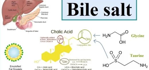
Bile salts & gall bladder function, Factors affecting gall bladder evacuation (Cholagouges)
Pancreas function, anatomy, parts, relations, ducts & blood supply