Histological structure of the small intestine, Mucosa, Submucosa and Intestinal glands
The small intestine connects the stomach and the large intestine, It is about 20 feet long and folds many times to fit inside the abdomen, It has three parts: the duodenum, jejunum, and ileum, It helps to digest food coming from the stomach, It absorbs nutrients (vitamins, minerals, carbohydrates, fats, proteins) and water from food so they can be used by the body.
Histological structure of the small intestine
The main function of the small intestine by its three parts; duodenum, jejunum, and ileum is the completion of digestion as well as absorption. Therefore, the small intestine has several histological modifications to fulfill its function, including:
1. The small intestine is the longest segment of the digestive tube, thus allowing prolonged contact between food and digestive enzymes to complete the process of digestion and to permit absorption of the digested products.
2. Modification of the luminal surface, 3 types of modification of intestinal mucosa and submucosa to increase the total surface area available for absorption of nutrients by 400 to 600 folds, these are:
- Plicae circulares (valves of Kerckring): grossly, the mucosa and submucosa of the small intestine show a series of permanent circular folds, These plicae are most developed in the jejunum.
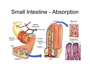
- Intestinal villi: these are mucosal outgrowths that project into the intestinal lumen. Each villus has a core of vascular lamina propria and is covered by simple columnar absorptive epithelial cells (the enterocytes) and goblet cells. In the duodenum, the villi are relatively short, broad, and leaf-like. In the jejunum, they are the longest and finger-like; whereas in the ileum they are short and finger-like. Accordingly, food absorption is maximal in the jejunum and is the least in the ileum.
- Microvilli: in the form of numerous closely-packed microvillous borders in the apical “luminal” cell membrane of the enterocytes covering the intestinal villi with its characteristic glycocalyx cell coat.
Histological organization of the small intestine
The three small intestinal segments have many common features except for certain histological structures characterizing each part, The wall of the small intestine is formed of the following layers:
Mucosa
It consists of the intestinal villi and the intestinal glands (crypts of Lieberkühn) that are lying in the lamina propria. The openings of the intestinal glands are present between the bases of the villi. The lamina propria with the intestinal glands is limited by the muscularis mucosae. which is formed of inner circular and outer longitudinal layers of smooth muscle fibers.
I- Intestinal villi: as mentioned above, they are projections of the mucosa. The covering epithelium of the villus is continuous with that of the intestinal glands.
Each intestinal villus is formed of a central core of loose connective tissue, extending from the lamina propria. It contains fenestrated capillary loops, a blindly-ended central lymphatic channel (central lacteal) and a few smooth muscle fibers. The villous epithelium, formed of 4 types of cells, these are:
1. The columnar absorptive cells “enterocytes”:
- They are tall columnar cells with oval basal nuclei, covering the surface of the villi and also present in the superficial parts of the intestinal crypts.
- Their luminal surface has a prominent microvillous border, with a well-developed cell coat forming the site of activity of many digestive enzymes secreted in the small intestine.
- The neighboring enterocytes are held together by junctional complexes that seal the intestinal lumen from the lamina propria.
2. Goblet cells:
- They are distributed among the absorptive columnar cells covering the villi and are also present in the superficial parts of the intestinal crypts.
- They are less abundant in the duodenum but increase in number towards the ileum.
3. M cells (microfold cells):
- They are broad cells found among the intestinal epithelium of the ileum overlying the lymphatic collections of the Peyer’s patches.
- Each cell has a few, short microvilli (microfolds) while the basal cell membrane. shows deep pockets that are occupied by the lymphocytes and by the processes of macrophages extending from the lymphatic aggregations under the epithelium.
- M cells are antigen-sampling cells that play an important role in intestinal immunological function.
4. Enteroendocrine cells: they are more abundant in the basal parts of the intestinal crypts. Many enteroendocrine cells have been identified in the intestinal mucosa, such as S cells secreting secretin hormone and Mo cells secreting motilin hormone.
Il- Intestinal glands (crypts of Lieberkühn):
These are simple tubular glands that extend deep through the full thickness of the laming propria to open between the bases of the villi. Each crypt is lined by 5 types of cells these are:
- Few absorptive columnar cells.
- Goblet cells.
- Enteroendocrine cells.
- Stem cells: they are located in the basal half of the intestinal crypts, They are low columnar cells usually seen in mitosis, They proliferate and move either superficially to differentiate and replace the villous epithelium; or downwards where they differentiate into all types of cells lining the intestinal crypt.
- Paneth cells: they are similar to peptic cells of the fundic glands with strongly basal basophilic cytoplasm and apical eosinophilic zymogen granules. They are located in the base of the intestinal crypts. They secrete the antibacterial lysozyme which can break down the bacterial cell wall, and thus play important role in controlling the microbial flora in the intestines.
Ill- Lamina propria of intestinal mucosa
It is occupied by the intestinal crypts of Lieberkühn. In the ileum, the lamina propria contains the Peyer’s patches, which are characterized by the following:
They are aggregations of lymphoid nodules and loose lymphoid tissue as a part of the mucosa-associated lymphoid tissue (MALT). They are located on the side of the intestine lying opposite to the mesenteric attachment (anti-mesenteric).
They extend through the muscularis mucosae and appear also in the submucosa. The epithelium over these patches is usually lacking the villi (ironed mucosal appearance) and contains the M cells.
Peyer’s patches serve as an immunological barrier against the antigens in the lumen with the secretion of IgA antibodies by the plasma cells, released across the mucosal epithelial cells into the intestinal lumen.
Submucosa
In the duodenum, the submucosa of its proximal part is occupied by Brunner’s glands, which are mucous glands with their ducts open into the bottom of the intestinal crypts. They secrete thick alkaline mucus to protect the duodenal mucosa by neutralizing the gastric chyme. in the ileum, extensions of the Peyer’s patches are usually present in the submucosa.
Musculosa: composed of inner circular and outer longitudinal layers of smooth muscle fibers.
Serosa: the small intestine is covered by the visceral layer of the peritoneum (the mesothelium) continuous with that of the mesenteries.
You can download Science online application on Google Play from this link: Science online Apps on Google Play
Small intestine function, anatomy, parts, Arterial supply of the duodenum, midgut & hindgut
Pancreatic secretion composition, regulation, function, Differences between jejunum & ileum
Bile salts & gall bladder function, Factors affecting gall bladder evacuation (Cholagouges)


