Tissues types, Epithelial tissue features, Covering and Glandular Epithelium
Tissue can be used to diagnose & classify diseases, Tissues can identify that the patient has cancer, what kind of cancer (e.g., kidney, prostate), and the characteristics of cancer (e.g., Stage II, grade or subtype of lung cancer), Tissue can be used to monitor responses to treatments to determine if cancer is progressing or responding or what side effects of treatment are occurring.
Tissues
The tissue is a group of cells or fluid that can perform a specific job in the body such as cells in an organ like the kidney or heart or blood cells that carry oxygen to and waste materials from the cells in the body, The basic tissue is a group of similar cells specialized to perform a common function, these tissues do not exist as isolated units but rather in associations forming body organs and systems. The human body is composed of only 4 basic tissue types:
- Epithelial tissue (Epithelium); arises from the three layers (ectoderm, mesoderm, and endoderm)
- Connective tissue; arises from the mesoderm
- Muscular tissue; arises from the mesoderm
- Nervous tissue; arises from ectoderm.
Epithelium
The epithelium is a tissue that covers the body surfaces, lines body cavities, and forms glands.
General features of epithelial tissue
- It is composed of closely- aggregated cells with very little intercellular substance.
- The cells are somewhat regular in shape, they are held tightly together by a specialized intercellular junctional complex.
- The epithelium is avascular i.e. not penetrated by blood vessels, it is nourished by diffusion from capillaries of the underlying connective tissue.
- Epithelial cells are separated from the underlying connective tissues by the basement membrane.
- The epithelium is rich in nerve supply, it receives a great number of sensory nerve endings from the underlying connective tissue.
- The epithelium has a high renewal rate with a continuous process of degeneration and regeneration.
Calcification of epithelia
The epithelium is classified according to its structure and function into:
- Covering epithelium: as sheets of contiguous cells that cover or line the body surfaces.
- Glandular epithelium: as glands (secretory cells) which originate by invagination of surface epithelial cells
- Special types of epithelium
Covering Epithelium
Epithelial cells are classified according to:
A. The number of layers:
- Simple epithelium consists of only a single layer of cells in which all the cells rest on the basement membrane.
- Stratified epithelium is formed of two or more cell layers, where only the basal cell layer rests on the basement membrane.
B. The shape of cells:
- Squamous: flat cells with flattened nuclei.
- Cuboidal: cubical cells in which the height and width are similar. Cuboidal cells have central rounded nuclei.
- Columnar: in which the height is greater than width. Columnar cells have oval basal nuclei.
Simple Epithelium
1- Simple squamous epithelium is composed of one layer of squamous cells that rests on a basement membrane. A surface view shows that the cells have a polygonal outline. A side view shows that cells are extremely flattened with flattened nuclei.
Sites: It is characteristically thin to allow material transport across it.
- InBowman’s capsule of the kidney
- Lining the alveoli of the lung for the diffusion of gases
- The endothelium is the simple squamous epithelium lining of the blood vessels and lymphatic vessels to permit smooth passage of blood and lymph
- The mesothelium is the simple squamous epithelium lining the serous membranes e.g. pleura, pericardium, and peritoneum.
2- Simple cuboidal epithelium is composed of one layer of cubical cells with central rounded nuclei. It is relatively thick compared to the squamous variety.
Sites: It is present in areas where ion exchange is required.
- Lining the thyroid gland follicles
- Lining the kidney tubules.
3- Simple columnar epithelium is made up of a single layer of tall columnar cells, The nuclei are ovoid and close to the base of the cell, It has 2 types:
- Non-ciliated simple columnar epithelium: their free surface is smooth and may have microvilli. Sites: lining most of the digestive tract and the gallbladder.
- Ciliated simple columnar epithelium: the free surfaces of the columnar cells have many cilia. Sites: lining the uterus and the oviducts (the Fallopian tubes).
4- pseudostratified columnar epithelium: This type is essentially simple, composed of only one layer but it has a false appearance of stratified epithelium, it consists of crowded columnar cells so that all cells lie in contact with the basement membrane but they do not all reach the surface.
Those that reach the surface are tall columnar cell while the other cells which do not reach the surface are short and triangular, Several layers of nuclei, each lies at the widest portion of the cell; giving a false appearance of stratification, This type of epithelium has non-ciliated and ciliated types.
Sites:
- Non-ciliated pseudostratified columnar epithelium: lining the male genital tract e.g. epididymis.
- Ciliated pseudostratified columnar epithelium: lining most of the respiratory system.
Stratified Epithelium
The stratified epithelium has a protective function and can withstand wear and tear than simple epithelium, It is multilayered composed of a basal cell layer of columnar cells, lying next to the basement membrane, Above it is one or more intermediate layers of polygonal cells, the shape of the free surface layer of cells determines the name of the stratified epithelium into:
1- Stratified squamous epithelium: The most superficial cells are flat squamous, this type of epithelium is either keratinized or non-keratinized.
- Keratinized stratified squamous epithelium: the superficial layers of this epithelium are composed of flat scale-like cells whose nuclei and cytoplasm are replaced with keratin, thus form a dry tough layer. Site: epidermis of skin covering the whole body.
- Non-keratinized stratified squamous epithelium. Sites: the wet surfaces subjected to wear and tear e.g. the mouth cavity, esophagus, and cornea.
2- Stratified cuboidal epithelium: The superficial cells are cuboidal. It is uncommon type, lining ducts of sweat glands.
3- Stratified columnar epithelium: The superficial cells are columnar, It is also an uncommon type of epithelium.
4- Transitional epithelium: This type of epithelium varies greatly in appearance i.e. it can change its shape and number of layers, according to the functional state of the organ.
Sites: lining the urinary passages; the ureter and the urinary bladder, In these sites, the lumen is subjected to volume changes due to emptying and distension, in the empty condition, the epithelium consists of several layers:
- A basal cell layer is formed of low columnar cells.
- A varying number of intermediate polygonal cell layers of which the cell layer immediately below the surface layer is pear-shaped.
- The surface layer is composed of large cuboidal single or binucleated, dome-shaped cells (umbrella cells) with a convex upper surface and a concave lower surface, the dome-shaped cells are so large that each cell covers about three of the underlying pear-shaped cells.
In the full (distended) condition, the epithelial cells glide on each other and become formed of only two layers:
- The superficial large flattened cells.
- The basal cuboidal cells.
Glandular Epithelium
This type of epithelium is formed of secretory cells that produce Secretion, which is sometimes stored as secretory granules. Glands arise from the covering epithelium which proliferates into the underlying connective tissue, glandular epithelium can be classified according to various criteria:
1- According to the number of cells:
- Unicellular glands consist of one cell e.g. goblet cells present in the lining of the small intestine and the respiratory tract.
- Multicellular glands consist of clusters of cells e.g. most glands of the body.
2- According to the presence or absence of a duct system:
- Exocrine glands, in which the secretion is carried by ducts e.g. salivary glands.
- Endocrine glands or ductless glands, in which the secretion is released directly to the blood vessels e.g. thyroid gland and suprarenal gland.
- Mixocrine (mixed exocrine and endocrine) glands contain the two types e.g. pancreas.
3- According to the mode of secretion (the secretory mechanism):
- Merocrine glands, in which the secretory granules are discharged by exocytosis through the cell membrane without losing any part of the cell e.g. pancreas, salivary glands, and goblet cells.
- Apocrine glands, in which the secretory granules produced is discharged together with the apical parts of the cytoplasm e.g. lactating mammary gland.
- Holocrine glands, in which the secretion produced is shed off with the whole-cell leading to its complete destruction e.g. sebaceous gland.
4- According to the nature of secretion:
- Mucous glands, which secrete a viscid glycoprotein secretion e.g. goblet cells and sublingual salivary glands.
- Serous glands, which secrete a watery secretion rich in enzymes e.g. parotid salivary gland.
- Mixed glands, which secrete both mucous and serous secretions e.g. submandibular salivary gland.
- Glands with special secretions e.g. ceruminous glands which secrete ear wax and sebaceous glands which secrete a fatty secretion (sebum).
5- According to the shape of the secretory portion:
- Tubular, in which the Secretory units are elongated and tubular in shape.
- Alveolar (acinar), in which the secretory units are rounded.
- Tubuloalveolar (tubuloacinar), in which the secretory units have both tubular and alveolar parts.
6- According to the branching of the ducts and branching of the secretory portion; exocrine glands could be classified into:
- Simple glands, which have only one unbranched duct and one secretory unit.
- Simple branched glands, which have one unbranched duct and branched secretory units.
- Compound glands, which have a branched duct system as well as branched secretory units
Special Types of Epithelium
A. Neuroepithelium: the epithelial cells differentiate to act as nerve receptors.
Sites:
- The taste buds of the tongue.
- The organ of Corti in the ear.
- The retina of the eye.
B. Germinal epithelium: specialized type of epithelium carrying the function of reproduction and produce germ cells, It is present in the testis and ovary.
C. Myoepithelial cells: they are modified stellate epithelial cells that surround the secretory units (the acini) and the ducts of the glands, they contain myosin and actin myofilaments; hence, they can contract and squeeze the secretion from the glands.
Sites:
- Sweat glands.
- Salivary gland.
- Mammary gland.
Functions of epithelium
- The most important function of covering epithelial membranes is the protection against injuries, bacteria, chemicals, and water.
- Secretion by glandular epithelium.
- Absorption by the epithelial lining of the small intestine and renal tubules.
- Filtration by the epithelial lining of certain parts in the kidney to facilitate urine formation and in the lungs for gas diffusion.
- Reproduction by the germinal epithelium of the testis and ovary that are specialized in producing sperms and ova respectively.
- Contraction by the myoepithelial cells.
- Receptor function by the neuroepithelium specialized in the perception of special sense stimuli.
DNA Repair types, definition & importance, Protein Biosynthesis & Genetic Code
DNA replication steps & rules, DNA polymerase enzymes & RNA primer synthesis
Gene mutations types, causes, examples & Regulation of Gene Expression
Protein biosynthesis steps, site, importance, inhibitors & Protein maturation
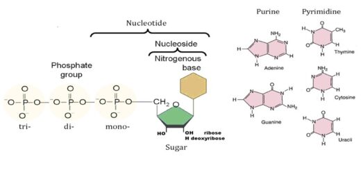
Importance of Nucleosides, Nucleotides, Purines, Pyrimidines & Sugars of nucleic acids