Common procedures of Interventional Radiology and the best Interventional Radiologists
Interventional Radiology (IR) procedures involve guiding a needle into the body using ultrasound or CT and accessing a particular organ (liver, kidney, stomach, etc) or a vessel (artery or vein), then guiding a wire and catheter through the needle into that area, Using a real-time x-ray camera. the wire and catheter can be guided through the body to perform whatever procedure is required.
Interventional radiology
Interventional radiology is the radiological subspecialty providing minimally invasive treatments performed under image guidance, As technology advances and high-quality imaging equipment becomes more widely available, Interventional radiology involves performing many imaging procedures to obtain images of the inside of the body.
The interventional radiologist interprets these images to diagnose injury and disease, and to perform many interventional medical procedures. Interventional radiology procedures are easier for the patient because they involve fewer incisions, less risk, less pain, and shorter recovery times, they depend only on local anesthesia, or anesthesia usually is not required.
Interventional radiologists
Interventional radiologists (IRs) are physicians who specialize in minimally invasive, targeted treatments performed using imaging guidance, Interventional radiology procedures are an advance in medicine that can replace open surgical procedures.

D. Mahmoud Abd ElAziz Ghalab is a famous interventional radiologist
The interventional radiologist is a medical doctor who has completed an accredited residency program in Radiology and is well-trained in different imaging equipment such as X-rays, magnetic resonance imaging (MRI), ultrasound, and computed tomography (CT) to diagnose disease. He can take the Doctorate in interventional radiology as a sub-specialty or enter a board exam given by the American Board of Radiology, and completes a fellowship-training program.
The interventional radiologist specializes in doing medical procedures that involve radiology, He can diagnose and treat diseases, and he can treat a wide range of conditions in the body by inserting various small tools, such as catheters or wires from outside the body, So, Interventional radiology can be used as a new alternative for some of the surgical procedures, and it can decrease the hospital stay and accelerate the recovery time for the patient.
Common IR Procedures
Interventional radiologists do many procedures, such as:
Needle biopsy: The Interventional radiologist puts a small needle into almost any part of the body, guided by imaging techniques, to take the tissue biopsy, This type of biopsy can offer a diagnosis without surgery, such as in breast biopsy, or biopsy from the focal hepatic lesion or suspicious lymph nodes.
Ultrasound-guided puncture to get vascular access: The interventional radiologist uses ultrasound to see the vascular structure either arterial or venous and determine the optimal site for puncture which minimizes any risk of bleeding or injuries to other structures, especially in difficult vascular access or patient with a bleeding tendency with the urgent need to insert CVC in ICU patients, Or as a preparatory step before any other procedures as venoplasty, peripheral angioplasty or Prostatic embolization.
Venous access catheters: The interventional radiologist puts the catheter into a large vein to give chemotherapy medicines (Port-a-cath), nutrition (PICC line), or hemodialysis (Permicath). This is determined according to the need of the patient and the strategy of management determined after multidisciplinary consultations between the IR consultant and other colleagues in different specialties, such as oncologists, nephrologists, and ICU specialists.
Injection of clot-dissolving medicines: The interventional radiologist injects clot-dissolving medicines such as tissue plasminogen activator, this medicine dissolves blood clots and increases blood flow to affected areas such as patients with recent cerebrovascular stroke, but this should be done within 3-6 hours of the attack of the ischemic insult.
Foreign body removal: The interventional radiologist puts the catheter into a blood vessel to remove a foreign body in the vessel using a snare.
Angioplasty: It comprises an X-ray of the arteries and veins to detect the problem in different peripheral vascular structures as a diagnostic step before the next step of management using balloon angioplasty to dilate a site of stricture, followed by stent insertion if needed to maintain adequate flow to the distal circulation and good perfusion with a minimally invasive procedure to get limb salvage technique, blockage or narrowing of the vessels, as well as other problems.
During the angioplasty: The Interventional radiologist puts a small balloon-tipped catheter into a blood vessel, Then he inflates the balloon to open up an area of blockage inside the vessel.
Embolization: The interventional radiologist injects different substances through the catheter into the blood vessel to stop blood flow through that vessel, This can be done to control bleeding or as management for malignant tumors such as primary liver hyper-vascular tumors such as hepatocellular carcinoma, or benign tumors as uterine fibroids or senile prostatic hyperplasia to allow their shrink in size and relief their pressure symptoms.
Gastrostomy tubes: The interventional radiologist puts a feeding tube into the stomach of a patient with mechanical obstruction due to esophageal neoplasm.
Stent placement: The interventional radiologist places a stent to overcome stricture in the biliary tree, venous stenosis, or arterial vascular insufficiency due to significant narrowing of a blood vessel at the site of a blockage, so he expands the stent to open up the blockage.
IVC filters: The doctor puts a small filter into the inferior vena cava (IVC), Which is a large vein in your abdomen, The filter catches blood clots that may go into your lungs to protect the patient from the risk of pulmonary embolism.
In cancer treatment: The Interventional radiologist gives the cancer medicine directly to the tumor site.
The best Interventional radiologists

D. Mahmoud Abd ElAziz Ghalab
Dr. Mahmoud Abd ElAziz Ghalab is a famous radiologist in Egypt, He earned a doctorate degree in Radio-diagnosis and intervention, the Faculty of Medicine, Alexandria University, Egypt, in cooperation with The University of Alabama in the USA, regarding the combination management of cases of Hepato-Cellular Carcinoma (HCC) using Trans-Arterial Chemo-Embolization (TACE) followed by Radio-Frequency ablation (RFA).
Dr. Mahmoud Abd ElAziz Ghalab is the head of the Intervention Radiology Department at Elekbal Hospital and Sahla Hospital in Alexandria since 2015. He is a co-founder of the Global Intervention Center (GIC) in Alexandria since (March 2016), and He is also a co-founder of the Radiology Department at Kafr Elsheikh University since (Feb-2016), He is a co-founder of the Intervention Radiology Unit in Kafr Elsheikh Main University Hospital since 2018.
Dr. Mahmoud Abd ElAziz Ghalab is the founder of the call scan company for portable radiology services in Alexandria and Damnhour since 2012, He is the founder of the first Intervention Radiology clinic in Kafr Elsheikh (Ghalab IR Clinic-GC) Since March 2020. D. Mahmoud Ghalab is a member of the first team for liver transplantation at Damanhour Teaching Hospital in El-Behera Government since March 2021.
You can contact him online and book at Dr. Mahmoud Abd ElAziz Ghalab
Email: [email protected]
Whatsapp number: +201003999893
IR procedures
Uterine fibroids: heavy menstrual bleeding and pain can be caused by benign tumors called fibroids, These can be treated by blocking blood vessels (uterine fibroid embolization, UFE) which leads to shrinkage, Embolization is combined with drug therapy (using small embolizing particles called embospheres or PVA) which targets the feeders of the tumor and preserving vascularity of the uterus itself.
Tumour therapies are intended to shrink or destroy tumours at their primary site or which have spread to other areas (metastases), This is an area of increasing interest and leads to improved survival with reduced morbidity. Liver, kidney, and other tumours (e.g. bone, lung) can be treated by destructive therapies (ablation) involving heat (radiofrequency, laser, microwave, focused ultrasound) or cold damage (cryotherapy), The treatment is performed and targeted using imaging machines (ultrasound, computed tomography or magnetic resonance imaging).
Gallstones are one of the most common upper abdominal disorders, Most are dealt with by laparoscopic surgery, When stones or tumors stop bile draining from the liver this causes jaundice, this is treated via a telescope passed down the throat (endoscopy) but in some cases, if there is contra-indication for endoscopy, so the patient referred for the interventional radiologist to perform drainage by placing catheter tubes through the biliary tree as a preparatory step to improve the patient general condition to allow stone removal later on.
Vascular Intervention (Arterial)
- Angioplasty/Stenting.
- Acute Arterial Catheter Directed Thrombolysis.
- Uterine Artery Embolisation – for Uterine Fibroids.
- Prostate Artery Embolisation – For Benign Prostatic Hypertrophy (BPH).
- Embolization for Post-Partum Bleeding.
- Embolization for Gastrointestinal Bleeding.
- Embolization for Trauma with liver laceration, splenic laceration, or renal lacerations.
- Endovascular Aneurysm Repair (EVAR).
Vascular Intervention (Venous)
- Varicocoele Embolisation.
- Long-Term Venous Access.
- Arterio-Venous Fistuloplasty.
- Thrombolysis.
- Thrombectomy.
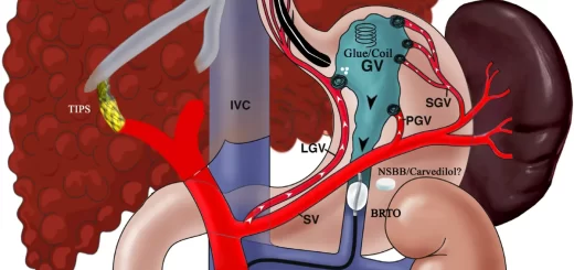
- Transjugular Intrahepatic Portosystemic Shunt (TIPS).
- Inferior Vena Cava (IVC) Filters.
Non-Vascular Intervention
- Nephrostomy & Ureteric Stent.
- Percutaneous Transhepatic Cholangiogram and Drain insertion (PTC- PTD).
- Radiologically Inserted Gastrostomy (RIG).
- Vertebroplasty.
- Image Guided Biopsies.
- Image Guided Drains.
- Cholecystostomy.
Interventional Oncology
- Interventional Oncology.
- Percutaneous Tumour Ablation.
- Portal Vein Embolization (PVE).
- Pre-Operative Tumour Embolization.
- Trans-Arterial Chemo-Embolization.
- Radio-embolization (Y90 – embolization).
You can download Science Online application on Google Play from this link: Science online Apps on Google Play
Interventional radiology types, Robotic endovascular systems advantages & disadvantages
Cone beam CT vs. Fan beam CT, CBCT advantages, disadvantages & uses
Artificial intelligence in radiology decision support systems, Medical Imaging & Healthcare
Enlarged prostate symptoms, Prostatic artery embolization (PAE) importance & risks
Hepatic Artery Embolization, Importance & risks of Embolization therapy for Liver cancer
Thyroid nodules, Thyroid Fine Needle Aspiration Biopsy importance & risks
Uterine fibroid embolization risks, importance & Minimally invasive procedure specialists