7 Powerful Facts About Neck Mass Symptoms, Types & Treatment in 2026
The neck connects the head to the torso and contains vital structures from several systems. A neck mass refers to any abnormal lump or swelling in the neck. These can range from benign (non-cancerous) to malignant (cancerous) and may be caused by a variety of conditions. Advanced neck mass treatment options in 2026 now allow earlier diagnosis, minimally invasive procedures, and improved recovery rates for both benign and malignant neck tumors
Neck Mass
A neck mass can appear as a lump, swelling, or enlarged area in the neck and may affect people of all ages. Some neck lumps are harmless and temporary, while others may indicate infections, cysts, thyroid disorders, or even cancer. Understanding neck mass symptoms and the difference between malignant and non-malignant neck lumps is important for early diagnosis and successful treatment.
Many people ignore painless neck swelling until it becomes larger or causes discomfort. In some cases, early medical evaluation can help detect serious conditions before complications develop. Modern diagnostic tools and treatment methods in 2026 have improved outcomes for both benign and cancerous neck masses.
What is a Neck Mass?
A neck mass is an abnormal lump or swelling located anywhere in the neck. It may develop suddenly or grow slowly over time. Neck masses can arise from lymph nodes, thyroid glands, salivary glands, muscles, tissues, or blood vessels. Some neck masses disappear after treating an infection, while others require imaging, biopsy, or surgical removal to determine the exact cause.
Classification of Neck Mass
Neck masses can originate from: Skin, Endocrine organs, Upper aerodigestive Tract, Vessels, or Lymph Nodes. They are classified into: Congenital and Acquired.
- Inflammatory.
- Benign Neoplasm.
- Malignant Neoplasm.
Evaluation
Evaluation, which leads to the proper treatment and the best outcome, follows the following 4 steps:
- Appropriate initial assessment.
- Role and technique of FNAB.
- Appropriate use and interpretation of imaging
- Management: The importance of specialized multidisciplinary care if malignancy is suspected.
Neck mass
Appropriate Initial Assessment
A careful history and examination can often help make the correct diagnosis of a lump in the neck. The clinical signs of size, site, shape, consistency, fixation to skin or deep structures, pulsation, compressibility, transillumination, or the presence of a bruit remain as important as ever.
- Age.
- Location.
- Risk Factors.
- Symptoms.
- Head & Neck Exam.
- General physical exam.
Age
- Children (Pediatric): Inflammatory, congenital, and Malignant-
- Young Adult: Congenital, Inflammatory, and Malignant.
- Adult (>40): Malignant, Congenital, and Inflammatory.
Location
Common neck mass locations include the following:
- Angle of mandible: parotid swelling.
- Lateral neck: Enlarged lymph nodes (LNs) – Central compartment: Thyroid swelling.
Role of 80% of neck masses
Cases of neck masses: 80% are neoplastic, of which 80% are malignant, of which 80% are metastatic.
Risk Factors
- Tobacco.
- Alcohol.
- HPV (Human Papiloma Virus) and HN (Head and Neck cancer).
- Male predominance in Cancer.
- Younger patients.
- Fewer traditional risk factors.
- Sexual behavior as a risk factor for multiple sexual partners (>6), higher rates of oro-genital contact with multiple partners.
- Sun Exposure, Ex, farmer.
Symptoms of Head and Neck Primary
- Otalgia, unilateral.
- Hemoptysis.
- Nasal obstruction (snoring).
- Unilateral hearing loss.
- Dysphagia.
- Epistaxis.
- Hoarseness.
- Sore throat.
Symptoms of Lymphoma
- Fever.
- Night Sweats.
- Weight Loss.
Physical Exam: What do we need to document?
- Location of the mass in the neck.
- Presence/absence of a primary in the head and neck.
- Presence/absence of generalized lymphadenopathy.
Non-neck mass
- Transverse processes of cervical vertebrae.
- Carotid bulb.
- Inferior belly of the omohyoid.
Role and Technique of FNAR
- Needle size 25 gauge.
- 12-15 Passes should be performed.
- Immediate assessment of adequacy by the Pathologist is the rule.
Fine Needle Aspiration Biopsy
Diagnosis of Lymphadenopathy
- Sensitivity 85-97%.
- Specificity 98-100%.
- Nondiagnostic 8-16%.
- Open Biopsy 22-30%.
Role of Open Lymph Node Biopsy
- Excisional/Incisional Biopsy may be necessary:
- Subclassification of lymphoma.
- Facilitate the diagnosis of poorly differentiated carcinoma.
- Persistently nondiagnostic FNAB.
IV Management: The importance of specialized multidisciplinary care cannot be emphasized more, particularly if malignancy is suspected:
- Benign
- Malignant
Non-malignant neck lumps
- Bening.
- Inflammatory.
1. Cystic hygroma (Lymphangiomas)
- It is a congenital lesion usually present within the first year of life. (Posterior Triangle).
- Usually remain unchanged into adulthood.
- Soft, cystic, multilocular, partially compressible, and brilliantly transilluminant, and may present with pressure effects.
- CT or MRI may help define the extent of the neoplasm.
- Treatment of Lymphangiomas includes injection with picibanil or excision for easily accessible lesions or those affecting vital functions.
- If it affects vital function and structure.
- Injection is very hazardous.
2. Branchial cleft cysts
- Remnant of branchial cleft (2nd).
- Most commonly occur in the second or third decades.
- Pain +/- (severe throbbing pain) (not infected = mild aching pain).
- Usually presents as a smooth, fluctuant non non-tender (tender), non-transilluminant mass mobile forwards and downwards, underlying the anterior border of the sternomastoid muscle.
- Branchial fistula or sinus (infection).
- Primary treatment involves controlling infection with antibiotics. followed by surgical excision.
3. Thyroglossal duct cyst
- This is a common congenital midline neck mass.
- Sometimes at the lateral edge.
- Pain and tenderness
- Can be moved transversally, but
- Elevates the protrusion of the tongue.
- Treatment is with initial control of infection with antibiotics, followed by surgical excision including the mid-portion of the body of the hyoid bone (Sistrunk’s procedure).
- Occasionally, these lesions become infected and resolve, or persist following drainage as a thyroglossal fistula.
4. Lipoma
- Lipomas are the most common benign soft tissue neoplasm in the neck.
- They are poorly defined, soft masses usually after the fourth decade.
- They are usually asymptomatic and soft.
- FNAC or an MRI Scan can confirm the diagnosis. MRI is the best.
- Surgery is indicated when the lump is increasing in size. cosmesis or when there is doubt about the accuracy of the diagnosis.
5. Sebaceous cysts
- These are common masses occurring often in older people, but can occur at any age.
- They are slow-growing, but sometimes fluctuant and painful when infected.
- Diagnosis is made clinically; the skin overlying the mass is adherent, and a punctum is often identified.
- Excisional biopsy confirms the diagnosis.
6. Cervical lymphadenopathy
Acute lymphadenitis
- Tender swelling.
- Antibiotic trial, less acute inflammatory nodes generally regress in size over 2-6 weeks.
- If the lesion does not respond, the biopsy is warranted.
TB cervical lymphadenitis
- Upper and middle deep cervical LN.
- Onset: gradually.
- Pain: +/-
- Systemic symptoms are unusual in the young.
- Abscess (painful, increase size, and skin discoloration).
- Mass: indistinct, firm, matted, fluctuate!
- Temperature! (Cold abscess) (hot if see infection).
- Treatment with anti-TB (6- 9 months) Rifampicin Ethambutol INH Pyrazinamid.
7. Carotid body tumor
- Rare tumor of chemo receptors) (40-60 years).
- Slow-growing, painless, sometimes pulsating lump may be bilateral.
- Side-to-side movement (not along the carotid).
- Symptoms of transient cerebral ischemia!
- Also known as Potato tumors (hard, non-tender).
- Palpation may induce a vasovagal attack.
- Biopsy is contraindicated: MRI (as its avascular mass).
- Angiography is the investigation of choice.
- Surgical removal is based on patient factors and presenting symptoms.
8. Pharyngeal pouch
- Diverticulum of the pharynx through the gap between the horizontal fibers of the cricopharyngeus muscle below and the lowermost oblique fibers of the inferior constrictor muscle above.
- History of froth and acid taste.
- Halitos is the regurgitation of food. There is no bile. or to it. (bad odor).
- Pressure on the swelling causes gurgling sounds and regurgitation.
- Treatment: cricopharyngeal myotomy.
9. Ludwig’s angina
- Rare but serious connective tissue infection of the floor of the mouth.
- Mostly due to dental infections.
- Signs of inflammation are present.
- Treatment: drainage of pus + antibiotic to cover aerobes with anaerobes.
10. Thyroid masses
- Thyroid neoplasms are a common cause of anterior compartment neck masses in age groups, with a female predominance, and are mostly benign.
- Fine needle aspiration of thyroid masses has become the standard of care, and ultrasound may show whether the mass is cystic.
- Unsatisfactory aspirates should be repeated, and negative aspirates should be followed up with a repeat FNAC and examination in 3 months.
Characteristics of malignant neck lumps
1. Lymphomas
- Painless lump: non-tender, smooth, and discrete.
- Slow-growing.
- Patient presented with malaise, weight loss, and pallor.
- Fever, rigor, and Hepatosplenomegaly.
- Mediastinal mass (SVC syndrome).
- Abdominal pressure on the IVC may cause bi lateral leg oedma.
- Other lymph nodes in the axilla, groin, and abdomen should be examined.
- Treatment: according to stage (radiosensitive).
2. Metastatic Lymph Nodes (commonest)
- Upper cervical lymph nodes (upper aero-digestive tract).
- Accessory chain of nodes in the posterior triangle (Nasopharyngeal malignancies).
- (Occult primary) The most common sites are the tonsil, base of tongue, nasopharynx, and the Pyriform sinus.
- Virchow’s LN (Troisier ‘s sign), abdominal and thoracic malignancies.
- Painless, non-tender, and hard masses.
- Work up: Search for the primary and deal with it.
Common Neck Mass Symptoms
Symptoms vary depending on the cause and whether the lump is benign or malignant.
Symptoms of a Neck Mass Include:
- Swelling or lump in the neck.
- Pain or tenderness.
- Difficulty swallowing.
- Hoarseness or voice changes.
- Persistent sore throat.
- Ear pain.
- Fever or infection symptoms.
- Weight loss without explanation.
- Enlarged lymph nodes.
- Breathing difficulty in severe cases.
Types of Neck Masses
- Congenital Neck Masses: These are present from birth and may become noticeable later in life. Examples: Branchial cleft cyst, Thyroglossal duct cyst, and Dermoid cyst.
- Inflammatory Neck Masses: These usually result from infections or inflammation. Examples: Swollen lymph nodes, Bacterial infections, Viral infections, and Tuberculosis-related lymphadenopathy.
- Benign Tumors: Non-cancerous growths that may enlarge slowly. Examples: Lipoma, Fibroma, Benign thyroid nodules, and Salivary gland adenoma.
- Malignant Neck Masses: Cancerous neck lumps may spread from nearby organs or originate in the neck. Examples: Lymphoma, Thyroid cancer, Metastatic head and neck cancer, and Salivary gland cancer.
Malignant vs Non-Malignant Neck Lumps
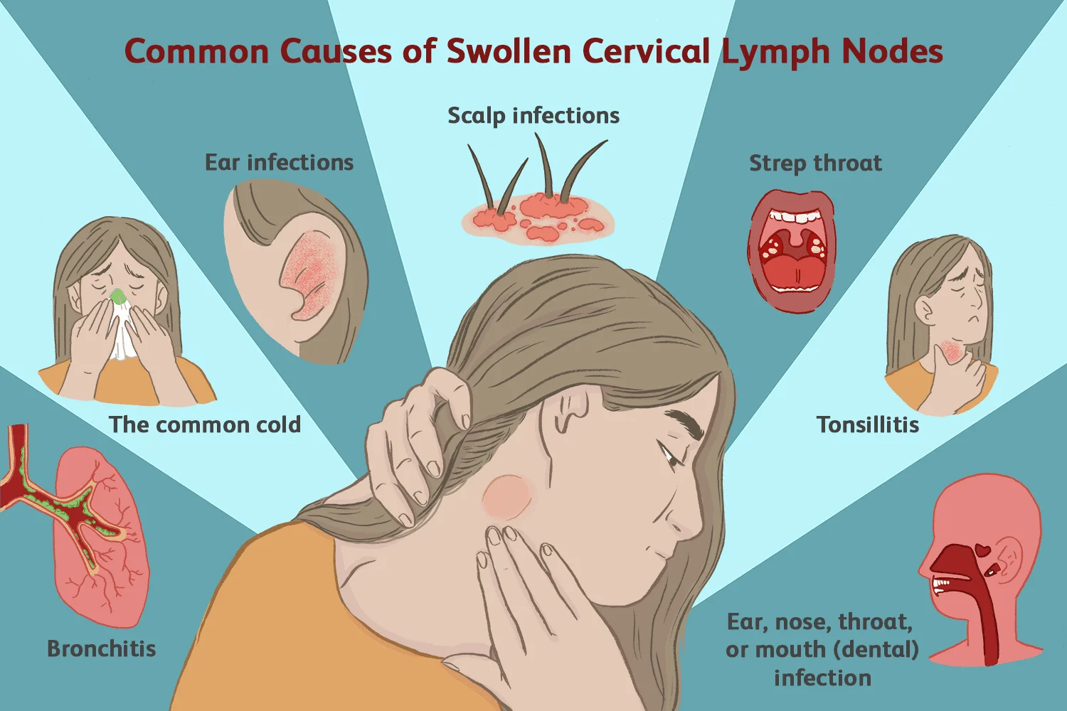
Non-malignant neck lumps are usually benign growths caused by infections, cysts, inflammation, or harmless tumors. These lumps are often soft, movable, and may become painful if linked to an infection. Swollen lymph nodes caused by colds, throat infections, or dental problems are among the most common non-cancerous neck masses. In many cases, benign neck lumps improve with medication or disappear once the underlying condition is treated.
Malignant neck lumps, on the other hand, are cancerous masses that may originate in the neck or spread from other parts of the body. These lumps are often hard, fixed in place, painless, and continue growing over time. Malignant neck masses may also be associated with symptoms such as unexplained weight loss, persistent hoarseness, difficulty swallowing, night sweats, or fatigue. Common causes include lymphoma, thyroid cancer, and metastatic head and neck cancers.
One of the biggest differences between malignant and non-malignant neck lumps is persistence. Benign lumps related to infections typically shrink within days or weeks, while cancerous neck masses usually remain and gradually enlarge. Doctors often use ultrasound, CT scans, MRI imaging, and fine needle aspiration biopsy to determine whether a neck lump is benign or malignant.
Early medical evaluation is essential because some malignant neck lumps may not cause pain during the early stages. Prompt diagnosis and treatment can significantly improve recovery rates and reduce complications. Anyone with a persistent neck lump lasting more than two weeks should seek professional medical assessment.
Causes of Neck Masses
Several conditions can lead to neck swelling or lumps.
Common Causes Include:
- Viral or bacterial infections.
- Enlarged lymph nodes.
- Thyroid disease.
- Salivary gland disorders.
- Cysts.
- Immune disorders.
- Benign tumors.
- Head and neck cancers.
Diagnosis of Neck Masses
Doctors use several methods to identify the cause of a neck lump.
Diagnostic Tests:
- Physical examination.
- Ultrasound imaging.
- CT scan or MRI.
- Blood tests.
- Fine needle aspiration biopsy (FNA).
- Endoscopy in some cases.
Early diagnosis helps determine whether the mass is benign or malignant and guides the best treatment approach.
Neck Mass Treatment Options
- Medications: Antibiotics or anti-inflammatory medications may treat infection-related neck swelling.
- Observation: Some benign neck masses only require monitoring.
- Surgical Removal: Cysts, benign tumors, or suspicious masses may need surgery.
- Cancer Treatment: Malignant neck lumps may require surgery, Radiation therapy, Chemotherapy, and Targeted therapy.
When Should You See a Doctor?
Seek medical attention if:
- The neck lump lasts more than two weeks.
- The swelling grows rapidly.
- You experience difficulty breathing or swallowing.
- There is unexplained weight loss.
- The lump feels hard or fixed.
- Hoarseness persists for weeks.
FAQ about Neck Mass
1. What are the first symptoms of a neck mass?
The first symptoms usually include a visible lump, swelling, tenderness, or enlarged lymph nodes in the neck.
2. Are all neck lumps cancerous?
No, many neck lumps are benign and caused by infections, cysts, or non-cancerous growths.
3. How can doctors tell if a neck lump is malignant?
Doctors use imaging scans, physical exams, and biopsies such as fine needle aspiration to determine if a neck mass is cancerous.
4. Can neck masses disappear on their own?
Some infection-related neck masses may disappear after treatment or with time, but persistent lumps should always be evaluated.
5. What is the best treatment for a neck mass?
Treatment depends on the cause and may include medications, observation, surgery, chemotherapy, or radiation therapy.
6. Is a painless neck lump dangerous?
A painless lump can sometimes indicate a malignant condition, especially if it persists or grows over time.
7. Can thyroid problems cause neck swelling?
Yes, thyroid nodules, goiter, and thyroid cancer can all cause neck masses or swelling.
8. When should I worry about a neck lump?
You should worry if the lump grows quickly, lasts longer than two weeks, causes swallowing difficulty, or is associated with weight loss or hoarseness.
Conclusion
Neck masses can range from harmless cysts to serious malignant tumors, making early diagnosis extremely important. Understanding neck mass symptoms, causes, and treatment options helps patients seek timely medical care and improve outcomes. If you notice persistent swelling or unusual neck lumps, consult a healthcare professional for proper evaluation and treatment.
You can subscribe to Science Online on YouTube from this link: Science Online
Cranial nerves types, Facial, Vestibulocochlear, Glossopharyngeal, Vagus, Accessory & Hypoglossal
Cranial nerves anatomy, function, Olfactory, Optic, Oculomotor, Trochlear, Trigeminal & Abducent
Triangles of the neck contents, Structure of Anterior & Posterior triangle of the neck
Facial muscles function, anatomy, arteries, veins, names & expressions
Skull function, anatomy, structure, views & Criteria of neonatal skull
Cranial cavity anatomy, function, structure, Dural folds, Cavernous sinus & Nerve supply of scalp