Differential diagnosis of abdominal masses 2026 & Is an abdominal mass always cancer?
An abdominal mass is an abnormal lump or swelling in the abdomen, which can arise from various organs, including the intestines, liver, kidneys, spleen, or reproductive organs. The cause of an abdominal mass can range from benign conditions to serious diseases, including cancer.
Causes of Abdominal Masses
Gastrointestinal Causes
- Hernia (inguinal, umbilical, incisional).
- Tumors (benign or malignant, e.g., colorectal cancer).
- Diverticular disease (diverticulitis with an abscess).
- Inflammatory bowel disease (Crohn’s disease with thickened bowel loops).
Hepatic (Liver) Causes
- Hepatomegaly (enlarged liver from fatty liver disease, hepatitis, or cirrhosis).
- Liver tumors (hepatocellular carcinoma, metastases).
- Liver cysts or abscesses.
Renal (Kidney) Causes
- Polycystic kidney disease.
- Renal tumors (renal cell carcinoma, Wilms’ tumor in children).
- Hydronephrosis (enlarged kidney due to obstruction).
Splenic Causes
Gynecological Causes
- Ovarian cysts or tumors.
- Uterine fibroids.
- Pregnancy-related masses (ectopic pregnancy, molar pregnancy).
Pancreatic Causes
- Pancreatic cysts or tumors.
- Chronic pancreatitis with pseudocyst formation.
Lymphatic Causes
Lymphadenopathy (enlarged lymph nodes due to lymphoma, tuberculosis).
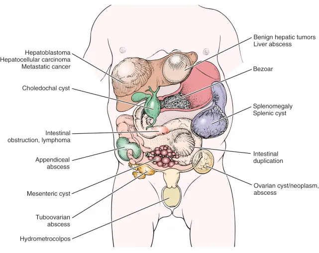
Abdominal masses
Symptoms Associated with Abdominal Masses
- Pain or tenderness.
- Swelling or bloating.
- Nausea and vomiting.
- Changes in bowel habits (constipation, diarrhea).
- Unexplained weight loss.
- Jaundice (yellowing of skin and eyes, if liver involvement).
- Fever (if infection is present).
Diagnosis
Doctors use a combination of: Physical Examination (palpation to locate the mass).
Imaging Studies:
- Ultrasound.
- CT scan or MRI.
- Blood Tests (tumor markers, liver/kidney function tests).
- Biopsy (if cancer is suspected).
Treatment
Treatment depends on the cause and may include:
- Surgical removal (e.g., hernia repair, tumor excision).
- Medications (antibiotics for infections, chemotherapy for cancers).
- Lifestyle changes (dietary modifications for liver or gastrointestinal conditions).
Differential diagnosis of abdominal masses
Abdominal Examination
1. History taking:
In patients presenting with a mass abdomen, generally, the following clinical features should be assessed carefully:
- Pain: Site, nature, aggravating or relieving factors, duration of pain, and referred pain.
- Vomiting: Type, content, hematemesis, relation to food, frequency.
- Jaundice: It is an important factor in relation to liver, gallbladder, or pancreatic masses.
- Bowel habits: Constipation, diarrhea, bloody diarrhea, furious diarrhea, tenesmus.
- decreased appetite and weight.
2. General examination
3. Local examination:
Position: Spine position
Exposure:
- Nipple to knee OR complete exposure of abdomen from xiphoid process to pubic or groin region.
- If it is embarrassing, cover the lower abdomen with the sheet.
Abdominal inspection
1. Abdominal shape & contour: (Normal – Scaphoid – Distended – Localized bulge).
A. Normal: Flat from xiphoid to pubis, and the umbilicus is at the center of the abdomen.
B. scaphoid abdomen: dehydration, cancer stomach, T.B. peritonitis).
C. distended abdomen:
- Localized (asymmetrical): huge organomegaly, tumors, operation, hernia.
- Generalized (symmetrical): 6F’s Fetus: pregnancy, flatus: gaseous distention, faces: constipation, fat: obesity, Fluid: ascites, fibroid.
2. Flanks: (Full Flanks, e.g., in ascites or empty flanks).
3. Epigastric pulsations: ask the patient to hold his breath.
4. Subcostal angle:
- Normal: right angle (70-110 degrees).
- wide angle (abnormal): Organomegaly (liver or spleen), ascites, COPD e.g. emphysema.
- Narrow angle (abnormal): Flat chest, visceroptosis, Pigeon chest.
5. Divercation of the recti: Ask patient to sit up in bed unsupported (raising up test).
- Due to a chronic increase in intra-abdominal pressure, leading to a weak abdominal wall.
- It is important to differentiate the abdominal wall from intra-abdominal masses:
- Abdominal wall mass: more prominent with tensing of abdominal wall musculature.
- Intra-abdominal mass will become less prominent or disappear.
6. Respiratory movements:
- Normal: Abdominothoracic in males & thoracoabdominal in females.
- Absent: In peritonitis.
- Paradoxical: In unilateral diaphragmatic paralysis.
7. Hernial arifices:
- Expansile impulse on cough due to a weak abdominal wall and increased IAP.
- Sites: Inguinal, umbilical, femoral, epigastric, incisional.
8. Umbilicus:
Site: normally midway between. Xiphisternum and symphysis pubis.
- Shifted upwards in pregnancy, ovarian mass, or tumor in the pelvis.
- Shifted downwards in ascites, hepatosplenomegaly, tumor in the upper abdomen.
Shape: normally inverted.
Everted in ascites and umbilical hernia.
Umbilical hernia: by Impulses on cough test.
Pigmentation around the umbilicus:
- Cullen sign: internal Hemorrhage leading to bluish discoloration of the umbilicus due to ruptured spleen, acute hemorrhagic pancreatitis, disturbed ectopic pregnancy, or malignant tumor with internal bleeding.
- Lymphoma: Brownish color.
Abnormal discharge from the umbilicus:
- Umbilical fistula: Fecal, urinary, biliary.
- Umbilical sinuses: abscess.
Dilated veins around the umbilicus: (Caput medusa).
Nodules around the umbilicus: Sister marry nodules: Indicate intra-abdominal malignancy due to metastasis.
9. Skin: Skin pigmentation: Cullen sign (periumbilical); grey turner sign (flanks), bluish discoloration denoting intra-abdominal hemorrhage.
Scars: Types of scars: splenectomy (meddle line), nephrectomy, cholecystectomy (right), hysterectomy (suprapubic), appendicectomy (right iliac). Impulses on cough or not (To exclude incisional hernia).
Striae: Due to the stretching of the abdomen.
- Striae alba (in obesity, pregnancy, ascites).
- Striae rubra (in Cushing, steroid uses, recent operation).
- Linea nigra (pregnancy line due to ⇑ melanin-stimulating hormone from the placenta).
Collateral (Dilated superficial veins): (dilated, elongated, tortious & congested).
Hair distribution: feminine hair distribution in liver disease.
scratch marks: in obstructive jaundice.
Petechiae & Purpura (red spots): due to platelets’ disfunction.
Ecchymosis & hematoma: due to coagulation disfunction.
10. Supra-pubic region: distended or not.
Distended in distended bladder or ovarian cyst.
11. Intestinal movements: Visible peristalsis denoting intestinal obstruction.
Abdominal palpation:
- Superficial Palpation: To gain the patient’s confidence and to detect local temperature, tenderness, parietal mass, or hypersthesia.
- Deep Palpation to feel the following:
If the patient is obese or rigid, use 2 hands to palpate; place one on the top of the other hand and feel with the lower hand.
If masses are felt, note the following:
- Inspection of the mass (6S): Site, size, shape, surface, skin overlying, special character.
- Palpation of the mass: Site, extent, surface, tenderness, consistency, movement with respiration, mobility, borders, plane of the swelling (by leg-raising test), and presence of other masses.
- Often, the mass needs to be examined for change of position: in sitting, in standing, in side position, after a brisk walk, in knee-elbow position for retro-peritoneal mass, and for puddle sign (but difficult to keep the patient in this position).
Percussion:
Percussion over the mass is important for predicting its anatomical location.
- If the mass has a dull note, then it is in the anterior abdominal wall, or in front of the bowel intra-abdominally, like liver, spleen, gall bladder (GB).
- If the mass is associated with an impaired resonant note, it arises from the bowel, such as the stomach, colon, or small bowel.
- If the mass is resonant on percussion, then it is probably in the retroperitoneal region.
- Other than this, liver dullness and free fluid in the abdomen should be elicited during percussion.
other:
- Per-rectal (PR) examination: It is done to look for any secondaries in a recto-vesical pouch, primary tumor, or relation of lower abdomen masses (pelvic masses).
- Per-vaginal (PV) examination: It is done to assess pelvic masses.
FAQ About Abdominal Masses
1. What is the differential diagnosis of abdominal masses?
Differential diagnosis of abdominal masses refers to identifying the possible causes of a lump or swelling in the abdomen. In 2026, doctors use physical examination, ultrasound, CT scans, MRI, blood tests, and advanced AI-assisted imaging to determine whether the mass originates from the liver, kidneys, intestines, ovaries, pancreas, or other abdominal organs.
2. Is an abdominal mass always cancer?
No, an abdominal mass is not always cancer. Many abdominal masses are benign (non-cancerous), such as cysts, enlarged organs, abscesses, fibroids, lipomas, kidney stones, or inflammatory conditions. However, some masses may be malignant, which is why proper medical evaluation and imaging are important for accurate diagnosis.
3. What are the common causes of abdominal masses?
Common causes include:
- Liver enlargement or liver tumors.
- Kidney cysts or hydronephrosis.
- Ovarian cysts or fibroids.
- Enlarged spleen.
- Pancreatic masses.
- Intestinal tumors.
- Hernias.
- Abscesses and infections.
- Benign fatty tumors (lipomas).
4. What symptoms can occur with an abdominal mass?
Symptoms may include:
- Abdominal swelling or bloating.
- Pain or discomfort.
- Weight loss.
- Nausea or vomiting.
- Loss of appetite.
- Changes in bowel habits.
- Fever in cases of infection.
5. How are abdominal masses diagnosed in 2026?
Modern diagnosis may involve:
- Ultrasound imaging.
- CT scan.
- MRI scan.
- PET scan for cancer evaluation.
- Blood tests and tumor markers.
- Endoscopy or colonoscopy.
- Image-guided biopsy.
- AI-supported diagnostic imaging systems.
6. Can abdominal masses be treated without surgery?
Yes, treatment depends on the cause. Some masses require medication, antibiotics, drainage procedures, or minimally invasive interventional radiology techniques, while others may need surgery or cancer therapy.
7. Are abdominal masses dangerous?
Some abdominal masses are harmless, while others may indicate serious diseases such as cancer, organ enlargement, or internal bleeding. Early diagnosis improves treatment outcomes and reduces complications.
8. What is the role of imaging in diagnosing abdominal masses?
Imaging helps determine:
- Size and location of the mass.
- Whether it is solid or fluid-filled.
- Relation to nearby organs.
- Signs of benign or malignant disease.
- Blood supply and tissue characteristics.
You can subscribe to Science Online on YouTube from this link: Science Online
Gastroesophageal Reflux Disease, Complications of GERD and Barrett’s oesophagus
Esophagus diseases, Dysphagia causes, Achalasia, and Symptomatic Diffuse Esophageal Spasm
Pharynx function, anatomy, location, muscles, structure, and Esophagus parts
Tongue function, anatomy, and structure, Types of lingual papillae, and Types of cells in taste bud
Mouth Cavity divisions, anatomy, function, muscles, Contents of Soft palate and Hard palate
Temporal and infratemporal fossae contents, Muscles of mastication and Otic ganglion